
Melanoma: causes and signs
Cutaneous melanoma is a neoplasm that originates from melanocytes, the cells responsible for the production of melanin
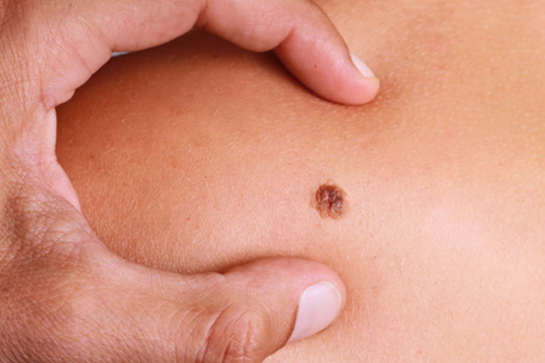
When it first appears, it may resemble an ordinary mole and go unnoticed by everyday household observation.
All moles can change over the course of a lifetime, but when these changes occur within a short period of time (ABCDE = Asymmetry, Border, Colour, Diameter, Evolution) it is important to consult your dermatologist and remove any doubts.
Today it takes only a few minutes to assess whether a mole is simply a mole.
Every dermatologist has an optical or digital instrument in his surgery that, thanks to a physical principle called epiluminescence, is able to visualise dermoscopic features not visible to the naked eye (e.g. network, globules, clumps, pseudopods, grey-blue veil, vessels, etc).
Epiluminescence dermoscopy is known by the popular term mole map and is performed periodically according to one’s risk profile.
There are people who are monitored more frequently (e.g. individuals with fair skin, red hair, blue eyes, who burn easily, with many moles or atypical nevi) and people who visit a little less often.
The frequency of check-ups is established at the time of the dermatological examination, based on a series of clinical and dermatoscopic factors that characterise the so-called nevus profile of the patient.
The causes of Melanoma
According to international scientific literature, repeated sunburn is associated with an increased occurrence of skin melanoma, although it can also occur in areas not exposed to the sun, with mechanisms that are still poorly understood.
Once a melanoma is identified by objective and dermatoscopic examination, it must be surgically removed, pending histological confirmation.
Today there is a tendency to remove the entire lesion for histological examination, instead of the more risky incisional biopsy if a melanoma is actually confirmed.
How widespread is skin cancer
Skin melanoma is a fairly widespread neoplasm and in Italy alone, thousands of new cases are registered every year (about 13 cases per 100,000 inhabitants).
There are populations with a higher incidence (e.g. light-skinned individuals from Australia), but also areas of the world where it is somewhat less common (dark-skinned individuals from countries in the equatorial belt).
What are the symptoms of Melanoma
To describe melanoma in simple words, we sometimes resort to the fairy-tale term of “ugly duckling”, meaning that after a while its features start to be so different from the rest of the patient’s other moles that its presence is already suspected in front of the mirror at home.
Other times these changes are less striking, slower and not always recognisable by the patient with the naked eye.
This is why it is important to periodically consult one’s dermatologist and have a clinical or instrumental confirmation of the degree of tranquillity of one’s mole.
The sudden bleeding of a mole, the sudden change in colour, borders, symmetry and size are already such an important signal to warrant a dermatoscopy.
How the diagnosis of Melanoma is made
The dermatologist is able to recognise a cutaneous melanoma from its clinical and dermatoscopic features.
There are a number of technical elements:

- thickened network
- chrysalis structure
- grey-blue veil
- vascular punctuation
- that allow the doctor to distinguish a simple melanocytic nevus from a melanoma that needs to be surgically removed.
At the time of the specialist examination, the dermatologist will check all skin neoformations, even those that are not pigmented, to search for and exclude a possible amelanotic melanoma (a neoformation that is clear as it lacks melanin).
The most common treatments for skin melanoma
Once a melanoma is recognised, it must be surgically removed, pending histological examination.
Immunotherapy is a fairly recent branch that is currently reserved by the oncologist for patients with melanoma in certain clinical conditions.
Prevention of Melanoma: advice
The best prevention remains the regular check-up of moles at one’s dermatologist.
With the outpatient instruments at his disposal, the dermatologist can also examine those atypical nevi that are not visible to the naked eye.
Read Also:
Emergency Live Even More…Live: Download The New Free App Of Your Newspaper For IOS And Android
Melanoma: Prevention And Dermatological Examinations Are Essential Against Skin Cancer
Epidermolysis Bullosa And Skin Cancers: Diagnosis And Treatment
Skin: What To Do In Case Of Folliculitis?
Childhood Psoriasis: What It Is, What The Symptoms Are And How To Treat It
Dermatological Examination For Checking Moles: When To Do It
What Is A Tumour And How It Forms
Rare Diseases: New Hope For Erdheim-Chester Disease
How To Recognise And Treat Melanoma
Moles: Knowing Them To Recognise Melanoma
Skin Melanoma: Types, Symptoms, Diagnosis And The Latest Treatments
Nevi: What They Are And How To Recognise Melanocytic Moles