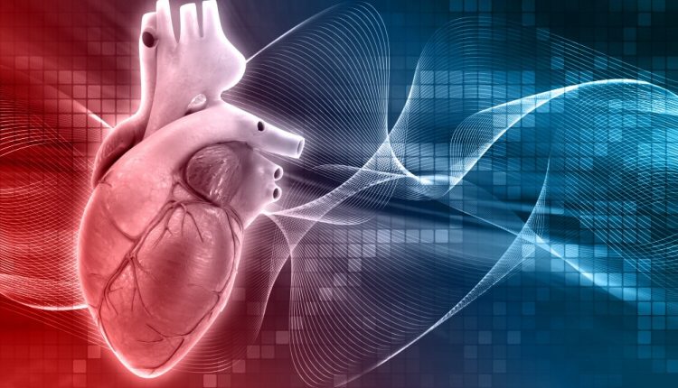
Coronary artery disease: ischaemic heart disease
Ischaemic heart disease is a condition that affects the coronary arteries: their progressive narrowing restricts the blood supply – and thus oxygen – to the heart
The main culprit of this medical condition is atherosclerosis, a condition characterised by the presence of atheromas (plaques with a high cholesterol content) at the level of the coronary wall, which can obstruct or reduce blood flow
The clinical manifestations of the condition are diverse, including myocardial infarction, which has a very high mortality rate.
What is ischaemic heart disease?
The term ischaemic heart disease is not used for a single pathological medical condition but encompasses a spectrum of situations that all have in common a reduced supply of oxygen to the myocardium compared to requirements.
The heart, requiring more oxygen than that carried by the coronaries, enters a state of distress, known as the hypoxic state.
But let us take a step back, starting with an analysis of the term.
‘Ischaemic heart disease’ is made up of two words, ‘cardiopathy’ meaning disease of the heart and ‘ischaemia’ meaning the diminution or suppression of the blood supply to a certain part of the body.
Tissues – in this case the heart muscle – affected by ischaemia are in a situation characterised by reduced oxygen supply (hypoxia or anoxia), but also by a reduced availability of nutrients that the blood carries.
The heart has a very high demand for oxygen and, when this is not met, there is a risk of damage and reduced cardiac function.
If, on the other hand, there is a total and sudden obstruction of the coronary arteries, this can lead to acute myocardial infarction, with the risk of circulatory arrest and thus death.
Undoubtedly the most frequent cause of ischaemic heart disease is atherosclerosis
A disease characterised by plaques (atheromas) that form in the wall of the blood vessels, preventing the proper flow of blood within the coronary arteries.
These atheromas, which have a lipid and/or fibrous composition, not only create a progressive reduction in the calibre of the coronary artery, but can also lead to ulceration of the artery walls, resulting in the risk of clots at the lesion and acute obstruction of the vessel.
In these cases, therefore, the risk of angina and myocardial infarction is very high.
Often, cardiac ischaemia is also due to coronary spasms, a medical condition that is much less frequent than atherosclerosis.
In addition to these medical conditions, there are also factors that definitely increase cardiovascular risk and could lead to ischaemic heart disease and these are:
- High cholesterol, due to congenital conditions or lifestyle habits. An excess of cholesterol in the blood definitely raises the risk of atherosclerosis.
- High blood pressure. Although it is often taken lightly, blood pressure is the first index to be considered and monitored.
- Diabetes. In the presence of diabetes, high cholesterol and hypertension, we could be facing a metabolic syndrome and therefore a clinical picture with a very high risk of cardiac ischaemia.
- Stress.
- Sedentary lifestyle.
- Obesity.
- Smoking
- Genetic predisposition.
As cardiac ischaemia encompasses a spectrum of conditions, the moment an imbalance is created between the heart’s need for substances and oxygen and the actual availability, various consequences, more or less serious, may occur.
This depends first and foremost on which vessel is occluded: if it supplies a very large part of the heart, the damage will be greater.
Other factors to be considered are the duration of the occlusion, the presence or absence of a collateral circle that could be created when the main vessel becomes blocked, and the general health of the person and the myocardium before the ischaemia.
Symptoms of ischaemic heart disease
There are, however, some common symptoms that occur with ischaemic heart disease: all of them or just some of them may occur; in any case, it is important to consult a doctor if we realise that we are not dealing with simple intercostal pain.
Certainly, chest pain will present itself, directly at the level of the heart (angina pectoris) but also at the mouth of the stomach, mistaken for reflux pain.
The pain may also radiate to the neck, jaw, left shoulder and arm.
You may experience severe breathlessness with difficulty in breathing, excessive sweating, nausea, vomiting and in some cases even syncope.
Is it possible to prevent it?
If for all diseases the best cure is prevention, this is particularly true for ischaemic heart disease.
We can start with a healthy lifestyle to keep our blood vessels and heart healthy by avoiding smoking and eating a balanced diet with low fat.
In addition, regular and constant physical activity as well as quitting smoking is a good idea.
If you realise that there is cardiac suffering or predisposing factors for ischemic heart disease, your doctor will prescribe certain drugs, such as aspirin and antiplatelet agents, to thin the blood; but also Beta blockers and Ace inhibitors to normalise blood pressure and heart rate.
The diagnosis of ischaemic heart disease passes through a series of instrumental tests, let’s see what they are
- We generally start with an electrocardiogram, which detects the first abnormalities that could indicate myocardial ischaemia.
- Holter. This is an ECG prolonged over 24 hours, used in cases of suspected angina.
- Stress electrocardiogram.
- Myocardial scintigraphy, which can consider blood flow both at rest and under stress.
- Echocardiogram, which allows a ‘snapshot’ of the heart and its functioning.
- Coronary angiography, to assess the health of the coronary arteries.
- CT scan of the heart, which can detect the presence of atherosclerotic plaques in the coronary vessels.
- Nuclear magnetic resonance imaging, which provides detailed images of the heart and blood vessels.
Complications
As mentioned earlier, there are several factors that determine the severity of ischaemia: in the most severe cases, cardiac damage is irreversible.
In fact, a heart cell can be without oxygen for between 20 minutes and 3 hours, after which it dies.
This cell necrosis is called an infarction, which becomes fatal if it affects a large number of cells.
These necrotised tissues do not regain their functionality, but become fibrous scar tissue, which is absolutely inert and therefore limits myocardial capacity.
The treatments used
Always talking about a broad spectrum of situations, we can generalise by saying that the goal of treating ischaemic heart disease is to restore proper blood flow to the heart muscle.
In less severe cases, this can be achieved with specific drugs; in worse cases, coronary revascularisation surgery will be necessary.
Let us start by explaining the pharmacological treatment.
Obviously, in this case in particular, there are no do-it-yourself treatments, but one must consult one’s treating physician who will work in collaboration with the cardiologist to establish the most appropriate treatment.
The following may be prescribed:
- Vasodilator drugs, such as nitrates and calcium channel blockers. Dilating the blood vessels, and thus also the coronary arteries, will ensure that the blood supply to the heart is sufficient for the muscle’s needs.
- Medications that thin the blood for proper circulation. We are talking in this case about anti-platelet aggregators.
- Medications that slow the heartbeat, such as beta-blockers. This will lower the blood pressure, reducing the work of the heart and thus the myocardium’s need for oxygen.
- Cholesterol control drugs, such as statins, to slow down or prevent the development and progression of atherosclerosis.
In some cases of more severe ischaemic heart disease, surgical intervention may become necessary. Two options are generally considered:
- Percutaneous coronary angioplasty. With this operation, a stent is inserted at the narrowing of the coronary artery during angiography. This reduces or completely eliminates the symptoms – but not the causes – of ischaemia. A stent is defined as a metal mesh that can be expanded to the exact size of the coronary lumen to be operated on.
- A coronary bypass, which is a much more invasive surgical procedure, may also be necessary. Vascular conduits are made to bypass the narrowed or occluded vessel.
Read Also
Emergency Live Even More…Live: Download The New Free App Of Your Newspaper For IOS And Android
Heart Valve Pathologies: Annuloplasty
Heart Valve Diseases: Valvulopathies
Anticoagulants: What They Are And When They Are Essential
Mitral Valve Narrowing Of The Heart: Mitral Stenosis
What Hypertrophic Cardiomyopathy Is And How It Is Treated
Heart Valve Alteration: Mitral Valve Prolapse Syndrome
Heart Rate Disorders: Bradyarrhythmia
Bradyarrhythmias: What They Are, How To Diagnose Them And How To Treat Them
Heart: What Are Premature Ventricular Contractions?
Life Saving Procedures, Basic Life Support: What Is BLS Certification?
Life-Saving Techniques And Procedures: PALS VS ACLS, What Are The Significant Differences?
Congenital Heart Diseases: The Myocardial Bridge
Heart Rate Alterations: Bradycardia
Interventricular Septal Defect: What It Is, Causes, Symptoms, Diagnosis, And Treatment
Supraventricular Tachycardia: Definition, Diagnosis, Treatment, And Prognosis
Ventricular Aneurysm: How To Recognise It?
Atrial Fibrillation: Classification, Symptoms, Causes And Treatment
EMS: Pediatric SVT (Supraventricular Tachycardia) Vs Sinus Tachycardia
Atrioventricular (AV) Block: The Different Types And Patient Management
Pathologies Of The Left Ventricle: Dilated Cardiomyopathy
A Successful CPR Saves On A Patient With Refractory Ventricular Fibrillation
Atrial Fibrillation: Symptoms To Watch Out For
Atrial Fibrillation: Causes, Symptoms And Treatment
Difference Between Spontaneous, Electrical And Pharmacological Cardioversion
‘D’ For Deads, ‘C’ For Cardioversion! – Defibrillation And Fibrillation In Paediatric Patients


