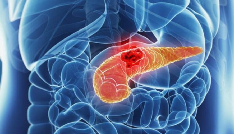
Diagnosis of pancreatic cancer: tests to be performed
Pancreatic cancer is by its very nature difficult to diagnose particularly in the early stages of the disease
More than half of all patients with pancreatic cancer have advanced disease and about a quarter already have regional spread.
There are many diseases that can mimic pancreatic cancer, such as an abdominal aortic aneurysm, ampullary carcinoma, intestinal ischaemia, gastric or pancreatic lymphoma, hepatocellular carcinoma (hepatoma), a stenosis or tumour of the choledocoel or endocrine pancreatic neoplasms.
Other situations such as acute pancreatitis, cholangitis, cholecystitis, choledochal cyst, chronic pancreatitis, gallstones (cholelithiasis), gastric cancer and peptic ulcer should not be forgotten.
Clinical approach: what investigations to do to diagnose pancreatic cancer
Laboratory data in patients with pancreatic cancer are generally not indicative, so it is necessary to rely on instrumental investigations, which may point more precisely towards a hypothesis of pancreatic neoplasia.
These investigations include:
- Computed tomography (CT)
- Transcutaneous ultrasound (ETC)
- Endoscopic ultrasound (EUS)
- Magnetic resonance imaging (MRI)
- Endoscopic retrograde cholangiopancreatography (CPRE)
- Positron Emission Tomography (PET)
The most difficult clinical situation in which to diagnose pancreatic cancer is in the patient with underlying chronic pancreatitis.
Indeed, in these cases all instrumental investigations may show morphological abnormalities that, particularly in the early stages, may not help to distinguish between a pancreatic carcinoma and chronic pancreatitis.
In many cases, tumour markers may also be of no help, as they appear elevated even during chronic pancreatitis.
In these patients, multiple modalities of instrumental investigations often have to be combined with close clinical follow-up with biopsy samples before a diagnosis of certainty can be reached.
Diagnosis of pancreatic cancer: laboratory findings
Laboratory findings in patients with pancreatic cancer are also generally non-specific.
Often, as in many cases of neoplasia, a normachronic anaemic state associated with thrombocytosis is observed.
But the most significant disorder is usually obstructive jaundice, manifested by an increase in bilirubin (conjugated and total), alkaline phosphatase, gamma-glutamyl transpeptidase and, to a lesser extent, aspartate aminotransferase and alanine aminotransferase.
Serum levels of amylase and/or lipase are elevated in less than half of patients with resectable pancreatic tumours and are elevated in only a quarter of patients with unresectable tumours.
However, about 5% of patients with pancreatic cancer have elevated amylase and lipase as a consequence of coexisting acute or chronic pancreatitis.
In the presence of liver metastases there is no clinical jaundice, but relatively low increases in serum alkaline phosphatase and transaminase levels may be present.
Patients with advanced pancreatic tumours and weight loss may also have general laboratory evidence of malnutrition (e.g. low albumin or cholesterol).
Tumour markers of pancreatic cancer
Carbohydrate antigen 19-9
CA 19-9 antigen is a protein present on the surface of some tumour cells and is most commonly found on circulating mucins in cancer patients.
It is also normally present in biliary tract cells and may be elevated in acute or chronic biliary tract diseases.
Approximately 5-10% of patients do not have the enzyme required to produce CA 19-9; in these patients with a low or absent CA 19-9 titre it will not be possible to monitor the disease with this tumour marker.
The significance limit for CA 19-9 is below 33-37 U/mL in most laboratories.
In the absence of biliary obstruction, intrinsic liver disease or benign pancreatic disease, a CA 19-9 value above 100 U/mL is highly specific for a neoplasm, usually pancreatic.
Evaluation of CA 19-9 levels has been used in addition to instrumental investigations to try to define the degree of resectability of pancreatic cancer, and in this context it has been shown that less than 4% of patients with a CA 19-9 level above 300 U/mL have resectable tumours.
Unfortunately, CA 19-9 is less sensitive for early-stage pancreatic carcinomas and therefore has not been shown to be effective for the early detection of pancreatic cancer or as a screening tool.
Although a standardised role for CA 19-9 in the diagnosis of pancreatic cancer has not been defined, it is of increasing importance in the staging and follow-up of patients with this disease.
Furthermore, during surgical, chemotherapeutic and/or radiotherapeutic treatment for pancreatic cancer, a declining CA 19-9 appears to be a useful surrogate result for clinical response to therapy. If biliary obstruction is not present, a rising CA 19-9 suggests progressive disease.
Preoperative CA 19-9 levels may be of prognostic value, with elevated levels indicating spreading disease with less chance of resectability.
Carcinoembryonic antigen (CEA)
Carcinoembryogenic antigen (CEA) is a high molecular weight glycoprotein normally found in foetal tissue.
It has been commonly used as a tumour marker in other gastrointestinal malignancies.
The reference range is less than or equal to 2.5 mg/ml.
Only 40-45% of patients with pancreatic carcinoma have elevated CEA values.
Since benign and malignant conditions other than pancreatic cancer can lead to elevated CEA levels, this marker is not a sensitive or specific indicator for pancreatic cancer.
Computed tomography
Because of its ubiquitous availability and its ability to image the entire abdomen and pelvis, abdominal CT continues to be the cornerstone of diagnostics used to evaluate patients with suspected pancreatic cancer.
New scanner models, using spiral CT scanning and dual- or triple-phase contrast enhancement, have significantly improved the sensitivity and specificity of the procedure.
On CT scan, malignant tumours appear as low-density lesions in relation to the surrounding structure and are often associated with pancreatic and/or biliary duct obstruction.
When lesions are visible, CT can also be used to perform targeted fine-needle biopsies and obtain a cytological/histological diagnosis.
Transcutaneous Ultrasonography
Although it is less expensive and generally more readily available than CT scan, transcutaneous ultrasound is less useful in pancreatic cancer than CT scan because the pancreas is often obscured by the presence of overlying gas from the stomach, duodenum and transverse colon.
However, ultrasound proves very useful as an initial screening test in the evaluation of patients with obstructive jaundice, highlighting intrahepatic or extrahepatic dilatation of the bile duct and identifying the site of obstruction.
Generally, a thoraco-abdominal CT scan, CPRE and/or magnetic resonance cholangiopancreatography should be performed to complete the diagnosis and perform an adequate staging of the disease.
Ecoendoscopy (EUS)
Echoendoscopy overcomes the physical limitations of standard ultrasound by placing a high-frequency ultrasonographic transducer on an endoscope, which is placed in the stomach or duodenum and this makes it possible to visualise the head, body and tail of the pancreas in detail.
Unlike CT scanning, the procedure requires conscious sedation, but due to the proximity of the pancreas to the EUS transducer, it is possible to perform a fine-needle cytoaspirate, which allows simultaneous and immediate cytological confirmation of pancreatic carcinoma at the same time as a pancreatic mass is detected.
EUS appears to be equivalent to dual-phase spiral CT scanning for assessing the degree of tumour resectability.
It is probably superior to computed tomography as a means of assessing the T-stage of the tumour, especially in defining portal vein involvement in the neoplastic lesion.
Endoscopic retrograde cholangiopancreatography (CPRE)
CPRE is an extremely sensitive means of detecting pancreatic and/or biliary ductal abnormalities in pancreatic carcinoma.
Among patients with pancreatic adenocarcinoma, 90-95% have imaging abnormalities, although they are not always highly specific for pancreatic carcinoma and may be difficult to differentiate from changes observed in patients with chronic pancreatitis.
CPRE is more invasive than the other available instrumental diagnostic modalities for pancreatic carcinoma and has a risk of pancreatic complications of approximately 5-10%.
For this reason, this investigation is nowadays usually reserved as a therapeutic procedure to resolve biliary obstruction and allow the therapeutic palliation of obstructive jaundice by the placement of a plastic or metal biliary prosthesis or to establish the diagnosis of unusual pancreatic neoplasms, such as intraductal mucinous neoplasms of the pancreas (IPMN).
CPRE particularly in the recent past has been used to diagnose pancreatic carcinoma cytologically/histologically by bile duct brushing (brush in the bile duct) or with biopsy forceps, although the diagnostic yield is less than 50 per cent.
Magnetic resonance imaging (MRI)
Interest in the use of magnetic resonance imaging continues to grow.
Dynamic, gadolinium-enhanced, 3D MRI can offer greater sensitivity in the detection of small pancreatic lesions as well as for the iconographic evaluation of the biliary tree and pancreatic duct.
Furthermore, MRI proves useful for more clearly defining the presence of liver metastases (particularly after chemotherapy), for resolving the suspicion of pancreatic neoplastic lesions, when CT scans are inconclusive, or in cases where patients are allergic to the contrast agents used with CT scans.
PET scan
PET scanning uses 18-F-fluorodeoxyglucose (FDG) to image primary tumour and metastatic disease.
PET scanning appears to be particularly useful in detecting occult metastatic disease.
Its role in the management of pancreatic cancer evaluation, however, is still under investigation.
False positive PET scans are not uncommon in the course of pancreatitis.
Needle biopsy
The need to obtain a cytological or tissue diagnosis of pancreatic cancer prior to surgery remains controversial and is highly dependent on the centre to which the patient is referred.
Arguments in favour of preoperative biopsy include its ability to provide evidence of pathology prior to surgery, to exclude unusual pathology, and to provide evidence of disease prior to the initiation of multidisciplinary treatment, such as neoadjuvant chemotherapy.
Arguments against preoperative biopsy of pancreatic lesions are that biopsy results normally do not alter therapy, that biopsy may cause neoplastic dissemination and interfere with definitive surgery.
Studies on the risk of peritoneal contamination with CT-guided biopsy have suggested that this risk is in reality very low.
EUS-guided fine-needle aspiration provides the additional advantage of aspiration through the tissue, which would still be included in the operative field should the patient undergo resection surgery.
Fine-needle aspiration under ultrasound or echoendoscopic guidance has proven to be the most effective means of making a definitive cytological diagnosis of pancreatic carcinoma in more than 85-95% of patients.
Three morphological features were identified in the cytology/histology analysis as being significantly associated with pancreatic cancer:
- Anisonucleosis
- Atypical single epithelial cells
- Mucinous metaplasia
The risk of neoplasia is indeed low when none of these 3 criteria is met, moderate when one factor is present, high when 2 or 3 of them are present.
The diagnostic yield of fine-needle cytoaspirate or CT-guided biopsy is around 50-85% in visible lesions.
Read Also
Emergency Live Even More…Live: Download The New Free App Of Your Newspaper For IOS And Android
Pancreatic Cancer: What Are The Characteristic Symptoms?
Gestational Diabetes, What It Is And How To Deal With It
Pancreatic Cancer, A New Pharmacological Approach To Reduce Its Progression
What Is Pancreatitis And What Are The Symptoms?
Kidney Stones: What They Are, How To Treat Them
Acute Pancreatitis: Causes, Symptoms, Diagnosis And Treatment
Pancreas: Prevention And Treatment Of Pancreatic Cancer
Acute Pancreatitis: What Is The Role Of Nutrition
Chemotherapy: What It Is And When It Is Performed
Ovarian Cancer: Symptoms, Causes And Treatment
Breast Carcinoma: The Symptoms Of Breast Cancer
CAR-T: An Innovative Therapy For Lymphomas
What Is CAR-T And How Does CAR-T Work?
Radiotherapy: What It Is Used For And What The Effects Are
Acute Pancreatitis: Causes, Symptoms, Diagnosis And Treatment
Gestational Diabetes, What It Is And How To Deal With It
Pancreatic Cancer, A New Pharmacological Approach To Reduce Its Progression
What Is Pancreatitis And What Are The Symptoms?
Kidney Stones: What They Are, How To Treat Them
Symptoms And Treatment For Hypothyroidism
Hyperthyroidism: Symptoms And Causes
Surgical Management Of The Failed Airway: A Guide To Precutaneous
Thyroid Cancers: Types, Symptoms, Diagnosis


