
Synovitis: definition, causes, symptoms and diagnosis of synovial membrane inflammation
It can happen that the synovial membrane – the tissue that lines the inner surface of the joint capsule – becomes inflamed
The reasons that lead to this process may be different, in each case we speak of synovitis.
There are acute forms as a result of trauma, infection, allergy or intoxication, but also chronic forms, which emerge as a result of degeneration of the joint cartilage.
Sometimes, synovitis is the consequence of certain dysmetabolic or rheumatic diseases, such as gout and rheumatoid arthritis, or tumours of the synovial membrane.
Let’s take a closer look at everything there is to know about this pathology, to recognise it and deal with it in the best possible way.
What is synovitis
As already mentioned, synovitis is the inflammation, acute or chronic, involving the synovial membrane, the portion of tissue that lines the inside of joints.
When the membrane becomes inflamed, it produces more synovial fluid, leading to swelling in the joint.
It may happen that synovitis also extends to cartilage and tendons, in which case we speak of tenosynovitis, or it may involve other structures adjacent to the synovium, in which case we speak of arthrosynovitis.
What is meant by synovial membrane and how the joint works
- As we have mentioned, synovial membrane refers to a thin portion of connective tissue in joints that internally lines the joint capsule, the articular portion of the bone, and all structures that are part of the joint, such as tendons and ligaments.
- Its specialised function is to produce synovial fluid, a fluid with a protective function and to remove all debris caused by wear and tear.
The term was coined by Paracelsus himself, comes from Latin and means egg: in fact, synovial fluid closely resembles egg white in colour and consistency.
Synovial fluid is contained within
- Synovial sacs: these have the specificity of cushioning any movement in the joint and reducing friction between the bones so that movement is smoother.
- Synovial sheaths: these structures line the tendons and reduce friction from rubbing.
- Symptoms common to all forms of synovitis are
- Swelling and swelling of the joint.
- Local pain that becomes more intense as the inflammation progresses. If action is taken late and the synovitis has become very severe, the synovial membrane may thicken to the point of eroding the bone, greatly increasing the pain.
- Joint effusion: due to inflammation, the membrane produces more synovial fluid than normal.
- Limitation of movements or even inability to perform some of them (e.g. extending the leg if the affected joint is the knee).
- Localised heat, caused by the inflammation, which can cause erythema (reddening of the skin).
In more severe forms, inflammatory nodules may appear that protrude into the joint cavity.
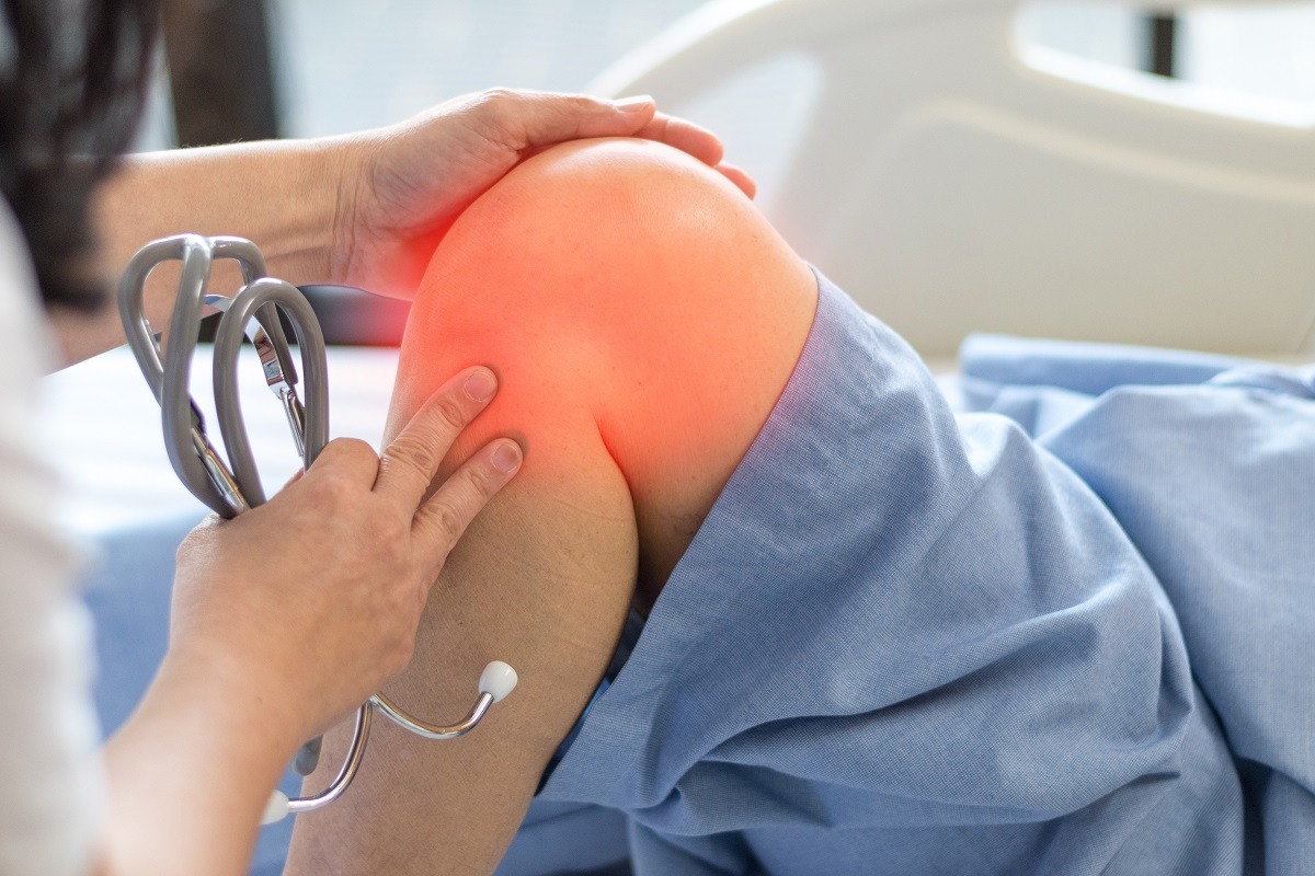
The joint most affected is usually the knee due to the increased stress it is subjected to, but it is possible for any other joint to be affected.
However, some of these symptoms – most of them – are common to other knee conditions: if synovitis is suspected, it is always a good idea to consult your doctor for a correct diagnosis.
The causes
As we have seen, the origin of synovitis can be ascribed to various factors, and among these are infections, trauma, allergies and intoxication.
In particular, acute forms of synovitis are caused by trauma or are secondary synovitis due to infectious diseases such as cerebrospinal meningitis, scarlet fever or typhus.
Acute forms may be exudative, i.e., inflammatory fluid infiltrates the joint cavity and mixes with synovial fluid.
Different are chronic synovitis, which may be
- Bacterial in nature, following particular diseases such as syphilis and tuberculosis.
- Consequent to particular conditions of degeneration or suffering of the joint, such as in patients suffering from arthrosis.
Diagnosis
Since the same symptoms can occur in different pathologies, it is impossible to self-diagnose this condition.
It is important, when experiencing the symptoms described above, to consult your doctor who will be able to direct you to the most suitable specialist, if necessary.
The medical diagnosis of synovitis will begin with an anamnesis, in which all the general information needed to understand the problem will be collected.
This is followed by an objective test, in which the doctor can detect the clinical manifestations of the problem.
Usually, diagnostic imaging tests such as X-rays, MRI, CT scans or arthroscopy may be prescribed to confirm the diagnosis.
Often, synovial fluid analysis is also recommended to rule out or confirm the presence of other pathologies that may cause synovitis, such as traumatic or rheumatoid arthritis, arthrosis or gout.
Treatments: conservative, local infiltrative and surgical
Depending on the severity and cause of the synovitis, the doctor will be able to recommend the best treatment to resolve the problem.
The first treatment recommended will probably be conservative treatment, which involves:
- Rest.
- Ice packs, applied regularly throughout the day.
- Use of an elastic bandage, as indicated.
- Administration of anti-inflammatory drugs in order to relieve painful symptoms.
- Taking certain supplements, which may support conventional therapies to counteract the symptoms of the disorder.
If conservative treatment does not have the desired effect, local infiltrative treatments are used: through injections of a certain drug or substance into the joint, the pharmacological action can be enhanced by concentrating the preparation locally.
Through injections it is possible – right from the first session – to reduce inflammation, slow down the worsening of the condition that the tissues were undergoing, and allow the patient to experience a reduction in pain.
The most commonly used drugs to perform what are commonly called infiltrations are corticosteroids, which are able to reduce the inflammatory response in the affected area.
Other substances that can be used are: hyaluronic acid, radioisotopes (able to penetrate synovial tissue without damaging cartilage, bone and other tissues) orgotein, some NSAIDs or glycosaminoglycans.
When even infiltrative treatment is not sufficient or in particular chronic conditions, surgery may be required.
The operation involves the total or partial removal of the inflamed or irreversibly damaged synovial membrane: the procedure is not at all invasive and in the days following the operation the patient quickly recovers normal mobility.
Generally, the operation is performed arthroscopically: although it does not allow for a complete synovectomy, it is decidedly non-invasive and allows for very rapid post-operative recovery times.
This is one of the most modern techniques and makes it possible to operate on joints and organs with minimal invasiveness: through small holes in the skin, the surgeon accesses the joint space with an arthroscope, a sort of miniature camera the size of a pen that allows the doctor to observe the affected area on a connected screen.
In this way, the orthopaedic surgeon will know how best to intervene: this procedure is therefore both diagnostic (it will allow the joint effusion and its nature to be clearly assessed) and therapeutic, because at the time of the arthroscopy, action can be taken to alleviate the symptoms or limit the damage observed.
Although, as we have seen, it is not a lengthy or invasive operation, there will be a recovery and convalescence time, in which it will be advisable to follow some useful advice and engage in proper rehabilitation.
First of all, it is a good idea not to immediately put body weight on the operated limb and, for a few days, depending on the doctor’s instructions, it may be necessary to use crutches for walking.
It will certainly be good to maintain the habit of ice packs two or three times a day.

Fundamental, as for many other operations on bones, muscles and joints, are physiotherapy sessions, usually lasting 2-3 months, necessary both to help recover the correct movements and to strengthen the muscles of the affected limb.
In any case, after surgery, the surgeon will make follow-up visits, during which he may perform hyaluronic acid infiltrations to improve the final result.
In the event that a synovial fluid test has revealed the presence of uric acid, it may be necessary to follow a special diet, and in the case of an established rheumatic disease, targeted therapies will have to be followed, recommended from time to time by the specialist.
Read Also
Emergency Live Even More…Live: Download The New Free App Of Your Newspaper For IOS And Android
De Quervain’s Stenosing Tenosynovitis: Symptoms And Treatment Of ‘Mothers’ Disease’ Tendinitis
Finger Twitching: Why It Happens And Remedies For Tenosynovitis
Shoulder Tendonitis: Symptoms And Diagnosis
Tendonitis, The Remedy Is Shock Waves
Pain Between Thumb And Wrist: The Typical Symptom Of De Quervain’s Disease
Pain Management In Rheumatological Diseases: Manifestations And Treatments
Rheumatic Fever: All You Need To Know
Arthrosis: What It Is And How To Treat It
Septic Arthritis: Symptoms, Causes And Treatment
Psoriatic Arthritis: How To Recognize It?
Arthrosis: What It Is And How To Treat It
Juvenile Idiopathic Arthritis: Study Of Oral Therapy With Tofacitinib By Gaslini Of Genoa
Arthrosis: What It Is, Causes, Symptoms And Treatment
Rheumatic Diseases: Arthritis And Arthrosis, What Are The Differences?
Rheumatoid Arthritis: Symptoms, Diagnosis And Treatment
Joint Pain: Rheumatoid Arthritis Or Arthrosis?
Cervical Arthrosis: Symptoms, Causes And Treatment
Cervicalgia: Why Do We Have Neck Pain?
Psoriatic Arthritis: Symptoms, Causes And Treatment
The Causes Of Acute Low Back Pain
Cervical Stenosis: Symptoms, Causes, Diagnosis And Treatment
Cervical Collar In Trauma Patients In Emergency Medicine: When To Use It, Why It Is Important
Headaches And Dizziness: It Could Be Vestibular Migraine
Migraine And Tension-Type Headache: How To Distinguish Between Them?
First Aid: Distinguishing The Causes Of Dizziness, Knowing The Associated Pathologies
Paroxysmal Positional Vertigo (BPPV), What Is It?
Cervical Dizziness: How To Calm It Down With 7 Exercises
What Is Cervicalgia? The Importance Of Correct Posture At Work Or While Sleeping
Lumbago: What It Is And How To Treat It
Back Pain: The Importance Of Postural Rehabilitation
Cervicalgia, What It Is Caused By And How To Deal With Neck Pain
Rheumatoid Arthritis: Symptoms, Causes And Treatment
Arthrosis Of The Hands: Symptoms, Causes And Treatment
Arthralgia, How To Cope With Joint Pain
Arthritis: What It Is, What Are The Symptoms And What Are The Differences From Osteoarthritis
Rheumatoid Arthritis, The 3 Basic Symptoms
Rheumatism: What Are They And How Are They Treated?
De Quervain Syndrome, An Overview Of Stenosing Tenosynovitis