
What is the peritoneum? Definition, anatomy and contained organs
The peritoneum is a thin, almost transparent, mesothelial serous membrane found in the abdomen that forms the lining of the abdominal cavity and part of the pelvic cavity (parietal peritoneum), and also covers a large part of the viscera contained within it (visceral peritoneum), while at the same time attaching them to the walls of the cavity (viscera ligaments)
The term peritoneum derives from the Greek περί (perì ) meaning around and τονείος (tonéios) meaning covered, which in turn comes from the verb τείνω (téinō), to cover: in fact, the peritoneum is the organ that covers around the organs of the abdomen and the abdominal wall.
The peritoneum is the largest of all serous membranes and, because of its arrangement, also the most complex
This complexity derives above all from the fact that instead of lining a single organ with a relatively uniform surface, as is the case with the pleura covering the lungs or the pericardium lining the heart, of which it is the abdominal equivalent, the peritoneum envelops several organs, arranged and oriented in the most varied ways and also having rather irregular shapes.
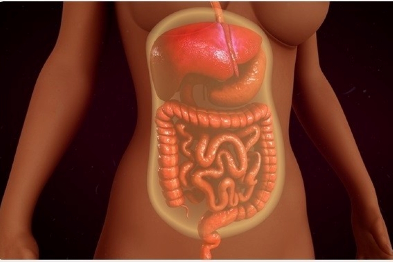
The visceral peritoneum, in accordance with this irregularity, also forms large folds between organs; a striking example is the large omentum, which stretches like an apron over the intestinal mass, starting from the large curvature of the stomach.
The peritoneum is made up of a superficial layer of mesothelial cells supported by thin layers of extraperitoneal connective tissue, which in certain regions is particularly rich in fat lobules, such as at the kidney, the inguinal region, certain duplications of the peritoneum and the outer surface of the large intestine; it appears that these fat accumulations perform a protective and supportive function for the organs. The peritoneum serves not only as a lining and support for the abdominal viscera, but also as a ‘conduit’ for the blood and lymph vessels and nerves of the abdominal region.
The peritoneum, like the other serous membranes, consists of a thin continuous lamina
Depending on its position in the abdominal cavity, it is distinguished into
- parietal peritoneum, the outermost layer, which lines the inner surface of the walls of the abdominal-pelvic cavity;
- visceral peritoneum, the innermost layer, which covers most of the viscera contained within the abdominal cavity.
Between these two layers there is a space, called the peritoneal cavity (or hollow), which is completely closed and is therefore a virtual cavity filled only with a small amount (about 50 ml) of a serous fluid that acts as a lubricant, allowing the two layers to slide together without excessive friction.
The visceral peritoneum, with its numerous folds around the abdominal organs, reduces the peritoneal cavity to a remarkably small, almost virtual space.
Some organs of the abdomen are completely enveloped by the peritoneum and are provided with a double leaflet, which is called the meso (e.g. mesentery for the small intestine, mesocolon for the colon, mesometrium for the uterus, and so on), that joins them to the parietal peritoneum of the abdominal wall.
In some cases, such as in the mesentery, a layer consisting of two welded sheets of visceral peritoneum tends to fuse with another sheet, giving rise to a fold that inserts itself into the posterior wall of the abdomen along an oblique line running from the duodenal-digiunal flexure to the right iliac fossa.
In other organs, such as the duodenum and the ascending and descending colon, the peritoneum forms an incomplete lining, leaving some uncovered areas in contact with the posterior abdominal wall.
The peritoneum is divided into two large regions, connected by the epiploic foramen
The large peritoneal cavity (or peritoneum of the peritoneal cavity proper).
The transverse mesocolon identifies:
- Supra-mesocolic space
- Submesocolic space, divided into two asymmetrical halves, right and left, by the mesentery. The right is smaller, closed at the level of the cecum, while the left sub-mesocolic space is open in the pelvis, divided from this by the mesosigma.
The omental bursa (or small peritoneal cavity)
One can distinguish:
- The Small omentum (gastrohepatic omentum or small epiploon) is connected to the small curvature of the stomach and the liver (via the ligaments: hepatogastric and hepatoduodenal, pars flaccida and pars densa respectively).
- The great omentum (or gastrocolic omentum or great epiploon or epiploic apron) originates from the visceral peritoneum that surrounds the posterior and anterior wall of the stomach, it starts from the great curvature of the stomach and descends like an apron in front of the loops of the small intestine to the theoretical line passing through the anterosuperior iliac crests, and then curves to form a loop anteroposteriorly and connects upward to the transverse colon, (4 leaflets in total); it performs the function of isolating and protecting the intestine.
Inguinal dimple
The inguinal dimples are compartments of the parietal leaflet of the peritoneum, which, resting on the transverse fascia, create dimples on the inner side of the anterior wall of the abdomen.
They are divided into:
- Outer inguinal dimple: this is located lateral to the inferior epigastric vessels.
- Mid inguinal dimple: lies between the inferior epigastric vessels and the lateral umbilical ligament (obliterated umbilical artery);
- Internal inguinal dimple: lies between the lateral umbilical ligament and the median umbilical ligament (obliterated urachus).
Classification of peritoneal structures
Structures located in the abdomen are classified as intraperitoneal, retroperitoneal or infraperitoneal based on whether they are actually covered by the visceral peritoneum and the presence or absence of mesenteries.
Intraperitoneal structures are usually mobile, whereas retroperitoneal structures are relatively fixed in their position.
Some organs, such as the kidneys, are defined as ‘primarily retroperitoneal’, while other organs, such as a large part of the duodenum and the pancreas (except for the tail, which is intraperitoneal), are considered to be ‘secondarily retroperitoneal’, meaning that these organs developed as intraperitoneal and later, with the loss of their meso, became retroperitoneal.
Pathologies
Like the other organs, the peritoneum is also subject to pathologies, which include acute or chronic, diffuse or circumscribed inflammatory processes (peritonitis, perivisceritis, abscesses), of a non-specific or specific nature.
Quite rare are primary tumours, such as fibromas, lipomas, myxomas, mesotheliomas, sarcomas, and secondary as a result of metastases from other organs.
Pneumoperitoneum, like pneumothorax in the thoracic cavity, is the presence of gas within the peritoneal cavity, which can occur in the event of perforations of the stomach or intestine; this creates a seriously dangerous situation, as accompanied by perforations there is often leakage of fluid from the stomach or intestine, which can cause a severe form of peritonitis.
Peritonitis is an inflammatory condition of the membrane and/or peritoneal cavity that occurs in cases of perforations or infectious outbreaks of the abdominal viscera, or both together.
It is a disease that leads to a severe clinical picture and often requires emergency intervention.
Ascites is an excess accumulation of fluid in the peritoneal cavity.

Adherent bridges are reactive fibrotic structures that lead to alterations in the normal anatomy and physiology of the small intestine.
Peritoneal dialysis
In a particular type of dialysis, called peritoneal dialysis, a solution is introduced by means of a catheter into the peritoneal cavity.
This fluid is left inside the abdomen for a certain period of time in order to absorb uremic toxins, which are then eliminated together with the solution through the catheter used beforehand.
This ‘cleaning’ takes place thanks to the large number of capillaries in the peritoneal membrane through the mechanism of molecular diffusion of substances.
Read Also
Emergency Live Even More…Live: Download The New Free App Of Your Newspaper For IOS And Android
Palpation In The Objective Examination: What Is It And What Is It For?
Acute Abdomen: Causes, Symptoms, Diagnosis, Exploratory Laparotomy, Therapies
Acute Abdomen: Causes And Cures
Peritonitis: Definition, Causes, Symptoms, Diagnosis, Types And Treatment
Abdominal Regions: Semeiotics, Anatomy And Contained Organs
Accumulation Of Fluid In The Peritoneal Cavity: Possible Causes And Symptoms Of Ascites
What Is Empyema? How Do You Deal With A Pleural Effusion?
Ascites: What It Is And What Diseases It Is A Symptom Of
Abdominal Health Emergencies, Warning Signs And Symptoms
Abdominal Ultrasound: How To Prepare For The Exam?
Abdominal Pain Emergencies: How US Rescuers Intervene
Abdominoplasty (Tummy Tuck): What It Is And When It Is Performed
Assessment Of Abdominal Trauma: Inspection, Auscultation And Palpation Of The Patient
Acute Abdomen: Meaning, History, Diagnosis And Treatment
Abdominal Trauma: A General Overview Of Management And Trauma Areas
Abdominal Distension (Distended Abdomen): What It Is And What It Is Caused By
Abdominal Aortic Aneurysm: Symptoms, Evaluation And Treatment
Hypothermia Emergencies: How To Intervene On The Patient
Emergencies, How To Prepare Your First Aid Kit
Seizures In The Neonate: An Emergency That Needs To Be Addressed
Abdominal Pain Emergencies: How US Rescuers Intervene
First Aid, When Is It An Emergency? Some Information For Citizens
Pain Management In Blunt Thoracic Trauma
Acute Hyperinflammatory Shock Found In British Children. New Covid-19 Pediatric Illness Symptoms?
Kidney Diseases, Kidney Ballot Manoeuvre: What It Is, How It Is Performed And What It Is Used For
Maneuver And Positive Or Negative Rovsing Sign: What Are They And What Do They Indicate?
Point Of Morris, Munro, Lanz, Clado, Jalaguier And Other Abdominal Points Indicating Appendicitis