
You have X-legs: discovering valgus knee together
Sufferers generally speak of ‘X-legs’. In reality, its medical name is valgus knee
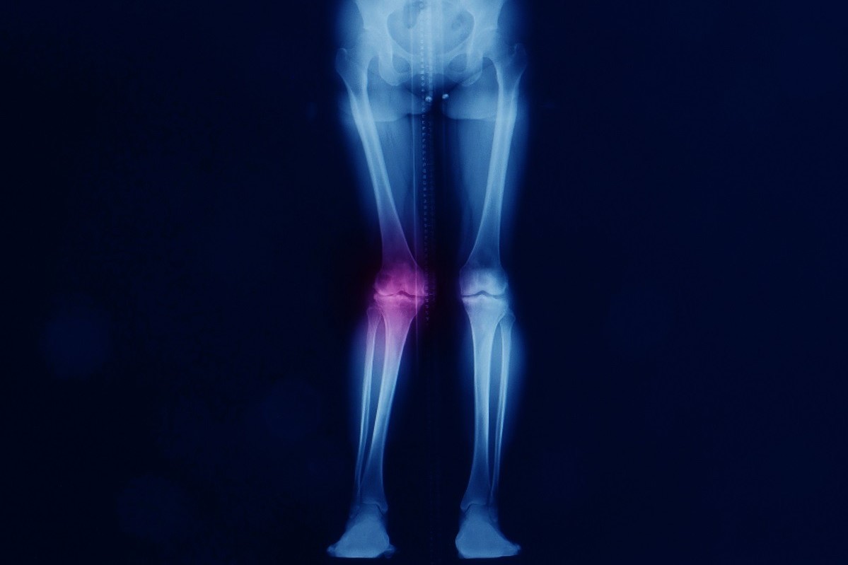
An anatomical deformity affecting the legs, valgus knee occurs when the knee deviates from the axis between the femur and tibia: as it descends towards the feet, the legs open outwards, thus forming an X-shape.
This is therefore the opposite phenomenon to varus knee, or ‘o-knee’, in which the legs form a kind of parenthesis with the knees pointing in two opposite directions.
Especially frequent in children, although it generally tends to resolve spontaneously by the age of 9, valgus knee can have very mild symptoms or lead to complications that compromise everyday mobility.
What is a valgus knee?
A valgus knee is a true anatomical deformity, although it is generally neither serious nor problematic.
As the name suggests, it is the knee, the synovial joint that connects the femur, kneecap and tibia, that is affected: the two knees point inwards, and the legs take on an x-shape.
Basically, there is a medial displacement of the knee, where ‘medial’ means ‘closer to the sagittal plane’ (the plane passing along the longitudinal axis of the body, which divides it into two symmetrical, mirror-like halves).
A valgus knee is usually bilateral, although in rare cases it may affect only one knee.
Knee valgus in children
Rather common in children up to 6 years of age, valgus knee is in the vast majority of cases a deformity that resolves spontaneously.
When the child approaches 7-9 years of age, thanks to the normal process of bone growth, the legs become straight again.
However, it is possible for the problem (especially if there are other cases in the family or if the child suffers from overweight or obesity) to persist.
What causes valgus knee in children is an imbalance in the activity of the growth cartilages.
These, located at the ends of the long bones (femur, tibia, humerus), allow them to lengthen in the course of growth through the production of new bone tissue.
If the cartilages lining the lower part of the femur and the upper part of the tibia grow out of balance, progressing faster on the inside than on the outside of the knee, valgus knee occurs.
If the child suffers from this (and realises it by sight alone, even before undergoing an X-ray of the pelvis and lower limbs), it is the paediatrician who will monitor the child to ensure that the deformity actually resolves spontaneously.
Especially when the disorder does not manifest itself in childhood but in adolescence, it is in fact possible that it worsens to the point of necessitating surgery.
In the case of a valgus knee in a child, it is good to know that, as a rule, nothing needs to be done (except to reduce body weight, in the case of obesity).
If, however, the disorder does not disappear, the specialist may prescribe partial epiphyseiodesis of the growth cartilage.
For all intents and purposes, this is a surgical procedure, to be performed during the developmental years, that temporarily blocks the growth cartilage of the femur and tibia in its inner part: a small plate is inserted, and the cartilage is forced to grow outwards only.
After about 12-18 months, when the alignment is complete, the plate is removed.
Bowed knee: the causes
In children, valgus knee has no cause: the growth cartilage simply does not function as it should.
In adults, on the other hand, there are causes that may also be co-present:
- the gluteal muscles are not as strong as they should be and the hips are weakened: if the gluteus does not push the hip outwards, the part of the femur included in it deviates inwards;
- excessive weakness of the quadriceps femoris or the hamstring semimembranosus and semitendinosus muscles, which are unable to perform an adequate pushing action;
- the person has a reduced capacity for dorsiflexion, i.e. to rise on the heels, and the foot goes into abnormal pronation;
- an anatomical predisposition, such as excessive pelvic width or an abnormality of the knee, femur, tibia, hip or foot.
A condition of obesity, rickets (newly formed bone tissue ossifies at the conjugate cartilage and areas of temporary calcification), a history of skeletal trauma or infection, or the presence of skeletal dysplasia (qualitative, morphological, and quantitative variation in the cellular structure of a tissue) is conducive to valgus knee.
Valgus knee: symptoms
The main symptom of a valgus knee is the particular conformation of the lower limbs, which take on an x-shape.
However, sometimes it can only be a cosmetic problem that is visible when the person is standing (more or less markedly) and especially when doing exercises such as squats and lunges.
Other symptoms that may appear are:
- pain in the kneecap or on the outside of the knee (due to inflammation of the iliotibial band, a band of collective tissue that runs down the side of the thigh ending just below the knee);
- unstable or poorly mobile knee, usually due to even minor strains or sprains of the medial collateral or anterior cruciate;
- gait abnormalities, which can also cause a rupture of the lateral meniscus.
When valgus knee is mild, it gives no symptoms and requires no treatment.
However, if it occurs in a severe form, action must be taken to prevent it from leading to complications such as gonarthrosis (knee osteoarthritis) or chondromalacia patella (pathology of the posterior cartilage of the kneecap, which causes inflammation).
Valgus knee: diagnosis and treatment
The orthopaedic specialist only needs a simple examination to diagnose valgus knee.
When the patient is standing, the knees tend to come together, the femurs descend obliquely in relation to the sole of the foot, and the tibiae progressively move away from each other.
If the doctor deems it appropriate, he or she may request an MRI scan that can give an indication of why the valgus knee has arisen.
If the knee valgus is mild, conservative therapy will be opted for, including the use of anti-inflammatory and chondroprotectants as needed, injections of hyaluronic acid, and physiotherapy and postural exercises: the former serve to strengthen the muscles that align the femur and tibia and to improve the elasticity of the ligaments, the latter counteract the overload on the knee.
If the patient is overweight or obese, losing kilos is necessary to prevent the disorder from worsening and leading to serious complications.

If the valgus knee is more ‘severe’, surgical intervention is necessary instead.
Younger patients, still in the development phase, undergo a minimally invasive operation that – thanks to a system of screws and plates – briefly interrupts the inward growth of the tibia and femur, without hindering outward growth.
It is an operation that involves a very short hospitalisation and equally short recovery times.
The adult patient, on the other hand, must undergo a complex operation: the femoral osteotomy
Generally practised in individuals under 60 years of age, and with a valgus knee greater than 10°, it is performed under selective spinal anaesthesia.
An incision is made on the lateral side of the distal femur and cut, and then a plate is inserted to fix the bone in its new position.
It is a real remodelling, not easy but with a very good prognosis.
Read Also
Emergency Live Even More…Live: Download The New Free App Of Your Newspaper For IOS And Android
Do You Suffer From Plantar Fasciitis? Don’t Worry, Here’s What It Is And How To Treat It
RICE Treatment For Soft Tissue Injuries
P.O.L.I.C.E. Vs R.I.C.E.: The Emergency Treatment For Acute Injuries
How And When To Use A Tourniquet: Instructions For Creating And Using A Tourniquet
Bone Callus And Pseudoarthrosis, When The Fracture Does Not Heal: Causes, Diagnosis And Treatment
First Aid, Fractures (Broken Bones): Find Out What To Look For And What To Do
Epicondylitis Or Tennis Elbow: How To Treat It?
Golfer’s Elbow, An Overview Of Epitrocleitis
Hip Pain: Causes, Symptoms, Diagnosis, Complications, And Treatment
Cartilage Injuries: Chondropathy
MOP Hip Implant: What Is It And What Are The Advantages Of Metal On Polyethylene
Hip Osteoarthritis: What Is Coxarthrosis
Why It Comes And How To Relieve Hip Pain
Hip Arthritis In The Young: Cartilage Degeneration Of The Coxofemoral Joint
Visualizing Pain: Injuries From Whiplash Made Visible With New Scanning Approach
Coxalgia: What Is It And What Is The Surgery To Resolve Hip Pain?
Elbow Fracture: What To Do After A Fall And Healing Time
Treating Injuries: When Do I Need A Knee Brace?
Wrist Fracture: How To Recognise And Treat It
Carpal Tunnel Syndrome: Diagnosis And Treatment
How To Put On Elbow And Knee Bandages
Knee Ligament Rupture: Symptoms And Causes
Lateral Knee Pain? Could Be Iliotibial Band Syndrome
Knee Sprains And Meniscal Injuries: How To Treat Them?
Stress Fractures: Risk Factors And Symptoms
Epiphysiolysis, An Overview Of This Pathology Of The Femur