
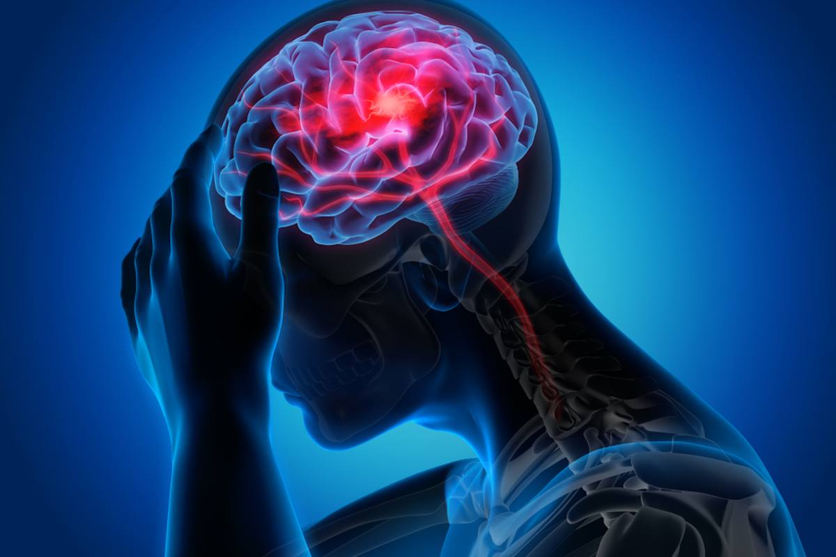
Brain damage: what is an ischaemic stroke?
An ischaemic stroke happens when a blockage cuts off the blood supply to part of your brain, killing brain cells. Damage to brain cells can affect how the body works. It can also change how you think and feel
A transient ischaemic attack (TIA or mini-stroke) is the same as a stroke but the symptoms only last for a short amount of time.
It is a major warning sign of a stroke and should always be taken seriously.
What happens when you have an ischaemic stroke?
If you have an ischaemic stroke, you will be given specialist care and treatment, including medication to reduce your risk of another stroke.
Afterwards, you will have support for your recovery including medical treatment and rehabilitation therapy.
The effects of your stroke depend on where the stroke was in your brain, and the amount of damage. For more information, see ‘Effects of stroke’ later on this page.
What causes an ischaemic stroke?
There are a number of reasons why blockages can form and cause an ischaemic stroke.
Atherosclerosis (narrowed arteries)
Atherosclerosis is where fatty deposits build up on the inside walls of the blood vessels carrying blood away from the heart (arteries).
These deposits are called plaques or atheromas.
Atheromas can build up in the large arteries in your neck leading to the brain, making them narrower and stiffer.
Atheromas can break down or become inflamed. When this happens a clot forms around the atheroma, which can block the blood vessel.
It may break off and move through the bloodstream into the brain, causing a stroke.
What causes atherosclerosis?
Some things can make you more likely to have a build-up of fatty materials in your blood vessels.
These include:
- Medical conditions including high blood pressure, high cholesterol and diabetes.
- Lifestyle factors such as smoking and being overweight.
- After a stroke, you should get advice about treating your medical conditions, and making healthy lifestyle changes.
Small vessel disease
Small vessel disease means having damage to the tiny blood vessels deep inside the brain.
The blood vessels become narrowed which reduces blood flow, and makes a stroke more likely.
It can also lead to many small strokes, as well as increasing the risk of bleeding in the brain.
Small vessel disease can be diagnosed on a brain scan, where it looks like scars in the brain structure.
It can affect your thinking ability and your mood, and it’s linked to cognitive decline and dementia.
What causes small vessel disease?
High blood pressure is a common cause of small vessel disease.
If you have high blood pressure, you will be offered treatment and advice for healthy lifestyle changes you can make to reduce your blood pressure.
Heart conditions
Atrial fibrillation (irregular heartbeat)
Atrial fibrillation (AF) means your heartbeat is irregular and may be abnormally fast.
Because the heart doesn’t empty itself of blood at each heartbeat, a clot can form in the blood left behind.
If this clot travels through the bloodstream to the brain, it causes a stroke.
AF often has no symptoms, but it can cause palpitations (feeling as if your heart is racing or skipping a beat).
Patent foramen ovale (PFO, or hole in the heart)
All babies in the womb have an opening between the right and left side of their heart, known as the ‘foramen ovale’.
This gap is needed while the baby is connected to the mother’s blood supply.
After birth, the baby’s blood circulation changes, and this gap usually closes up.
However, in as many as one in four people, the gap stays open.
This is known as a ‘patent’ (open) foramen ovale, or PFO. It’s sometimes referred to as a ‘hole in the heart’.
A PFO may be a risk for stroke, if a blood clot forms in the heart and travels up to the brain.
PFO does not always cause problems, and may not need to be treated.
In children, surgery is sometimes be used to close the PFO.
If you have a stroke, you will be assessed to decide if a PFO could have been a reason for your stroke, and what treatment you need.
Treatment options include blood thinning medication to reduce the risk of clots, or surgery to close the PFO.
Your doctor will talk to you about the best treatment for you.
Other heart conditions
Other heart problems, such as a recent heart attack or a mechanical heart valve, can also make a stroke more likely.
Arterial dissection (damage to the artery)
Arterial dissection is when the lining of an artery (a blood vessel leading away from the heart) gets torn.
It can happen after an injury, but it can also happen with no obvious cause.
Blood builds up in the damaged area, and a clot can form.
If this clot restricts the flow of blood to your brain, or moves up into your brain, it can cause a stroke.
Other causes
Sometimes stroke can be associated with other health conditions such as inherited blood clotting disorders or heart infections.
Your medical team will investigate these too.
How is an ischaemic stroke diagnosed?
If someone has any signs of a stroke, it’s time to call Emergency Number immediately.
Ambulance paramedics are trained in stroke.
They assess the person and take them to the right type of hospital for the treatment they need.
This could be a hospital with a specialist stroke unit or a hyper-acute stroke unit.
A stroke unit has an inter-disciplinary team of trained professionals who are experienced in stroke care.
The important thing when a stroke happens is time.
The faster someone can get to a specialist stroke unit, the better their chances of reducing damage to the brain.
Once you’re admitted to hospital, you have tests and checks to confirm if you have had a stroke, and what type of stroke it is.
THE WORLD’S RESCUE RADIO? IT’S RADIOEMS: VISIT ITS BOOTH AT EMERGENCY EXPO
Brain scan
If you have a suspected stroke, a brain scan should be carried out urgently, and if possible within one hour of arriving at hospital.
A brain scan can help doctors decide if you are suitable for an emergency treatment such as clot-busting treatment (thrombolysis) and mechanical clot removal (thrombectomy).
A computed tomography (CT) scan or a magnetic resonance imaging (MRI) scan is used to produce pictures of your brain.
Doctors use scans to rule out other causes of your symptoms, and see how much of your brain has been affected.
It also helps them decide how best to treat you, as treatments are different depending on the cause and timing of your stroke.
Some types of scan involve an injection to highlight the blood vessels of the neck and brain more clearly, known as computed tomography angiography (CTA) or magnetic resonance angiography (MRA).
Other checks and tests
Your blood pressure is checked, and you have blood tests for health conditions linked to stroke, such as diabetes and high cholesterol.
You may have other tests to check for conditions that could have contributed to your stroke.
These include an electrocardiogram (ECG), which checks for an irregular heartbeat, or a Doppler ultrasound scan to check for narrowing of the blood vessels in your neck.
How is an ischaemic stroke treated?
The main treatments aiming to break up or remove clots from the brain are usually only available within a few hours of a stroke.
But there is also a range of other types of care, including medication to reduce your blood pressure and reduce your risk of another stroke.
You will be monitored for signs of complications and given any treatment you need.
You will be assessed to find out how the stroke has affected you, and what help you need with your recovery.
Treatments to break up or remove clots
The two ways of treating clots in the brain are:
- Thrombolysis (clot-busting medication)
- Thrombectomy (mechanical clot removal)
Thrombolysis (clot-busting treatment)
Thrombolysis uses a clot-busting medicine to break up clots in the brain.
This helps to save more of the brain by allowing blood to return to the brain cells more quickly.
Fewer brain cells die, and the impact of the stroke can be reduced.
Thrombolysis needs to be given within four and a half hours of stroke symptoms starting.
In some circumstances doctors may decide that it could still be of benefit beyond four and a half hours.
Who can have thrombolysis?
This treatment is only suitable in around 12% of strokes, as there are guidelines for who can and can’t have it, to make sure it’s safe and effective.
To have thrombolysis, the person needs to reach hospital within the time limits for treatment (usually four and a half hours after symptoms begin).
If they don’t know when symptoms began, perhaps because the stroke happened while they were asleep, this may rule out thrombolysis.
Other reasons why thrombolysis can’t be given include:
- Your stroke was due to bleeding in the brain, not a clot.
- Your stroke is very mild.
- You have a bleeding disorder.
- You have recently had brain surgery.
- You have had another stroke or head injury within the past three months.
- Your current medication is not compatible with the clot-busting medication (alteplase).
If you are able to have thrombolysis, your medical team will explain the treatment to you.
You do not have to sign any paperwork – a verbal agreement is enough.
If you are unable to give your consent, either because of the effects of your stroke or another reason, the medical team will seek permission from your next of kin or another family member.
Time is critical so if it isn’t immediately possible to talk to your family, the medical staff will make the decision based on what they feel is in your best interests.
How it works
Thrombolysis uses a drug called alteplase, or recombinant tissue plasminogen activator (rt-PA).
You are given alteplase through a small tube into a vein in your arm.
During this procedure, which takes around one hour, the medical team will closely monitor your blood pressure, body temperature, breathing and blood sugar levels to ensure that they remain stable.
Risks of thrombolysis
Despite its benefits, there is a risk that thrombolysis can cause bleeding in the brain.
Within seven days of having thrombolysis, about one in 25 people treated will have bleeding in the brain, and this can be fatal in about one in 40 cases.
Doctors carefully balance the risk to the patient against the potential benefit of the treatment.
So someone may not be eligible for thrombolysis if they have conditions like internal bleeding or head injury, an aneurysm or uncontrolled high blood pressure.
Thrombectomy (clot removal)
Thrombectomy involves pulling the blood clot out of your brain using a clot retrieval device.
This is done by inserting a wire into a blood vessel in your groin, moving it up to your brain, and pulling the blood clot out.
Like thrombolysis, thrombectomy can help reduce brain damage by restoring blood flow in the brain.
This means that fewer brain cells die, lowering the chance of serious disability.

This procedure can be given to around 10% of people with ischaemic stroke.
It is only used when the clot is in a large blood vessel in the brain.
It should be carried out as soon as possible after the stroke and within six hours at the latest.
However it can be done up to 24 hours after the stroke, if doctors think it will benefit the person.
It’s often used in combination with thrombolysis (clot-busting medication).
What happens if the clot is not treated?
Clot removal and clot-busting treatment are effective at reducing disability after stroke, but only around 10-15% of people are able to have them.
These treatments are given on top of the standard stroke care, which includes tests, medication and therapy.
Without removal or clot-busting treatment, the blood clot usually breaks up naturally within a few days or weeks.
You are assessed to find out how the stroke is affecting you.
You will be supported to recover by specialist doctors, nurses and therapists working in a team to give you expert care.
You will also be given treatments to reduce your risk of another stroke, such as blood-thinning medications and pills for high blood pressure.
Surgery: decompressive hemicraniectomy
When the brain is injured the tissues can swell, just like a bruise.
If there is a lot of swelling, it can put pressure on other areas of your brain, causing further damage.
In a very small number of cases an operation may be needed to relieve pressure on your brain.
A decompressive hemicraniectomy involves opening up a section of your skull to allow the brain to swell outwards and relieve some of the pressure.
Treatments to reduce the risk of another ischaemic stroke
Medication
Most people who have an ischaemic stroke will be given blood–thinning medication to help prevent clots from forming.
For most people this will be a daily dose of aspirin followed by clopidogrel.
If you receive thrombolysis, you normally have to wait at least 24 hours before you can begin taking aspirin.
How long will I need to take blood thinning medication?
Most people will need to take blood-thinning medication for life.
There are two main types of blood-thinning medication, known as antiplatelets and anticoagulants.
Many people need antiplatelets such as aspirin and clopidogrel.
People with heart conditions like atrial fibrillation are more likely to have an anticoagulant such apixaban, dabigatran, edoxaban, rivaroxaban or warfarin.
Find out more about blood-thinning medications on our dedicated webpage.
Surgery for narrowed arteries in the neck (carotid artery disease)
Around 15% of ischaemic strokes are due to narrowed arteries in the neck, known as carotid artery disease.
This is diagnosed using specialist ultrasound scans of your neck.
Carotid artery disease is due to atherosclerosis, the build-up of fatty materials in your arteries.
Carotid artery disease is sometimes treated using a surgical procedure.
This means either removing the artery lining, or inserting a mesh cylinder (stent) to keep the artery open.
You’ll be assessed to decide on the best treatment to help reduce your risk of a stroke, which might include medication instead of surgery.
Your care in the first 24 hours after a stroke
The team on the stroke unit continue to monitor you closely for at least 24 hours to ensure you remain stable.
You should have a swallowing test within four hours of being in hospital, to make sure it’s safe for you to eat and drink, or take medicine by mouth.
You may see some signs of recovery from your stroke early on, but if you’re still showing lasting effects after 24 hours, you will have a full assessment with all the professionals on the stroke team.
The team can include physiotherapist, speech and language therapist, occupational therapist, dietitian, orthoptist and a psychologist.
After 24 hours, you will be supported to get up, or walk around if it is safe for you to do so.
If you’re not able to move about very much, the way you are positioned is very important to help you avoid problems with breathing, chest infections (pneumonia), shoulder pain or pressure sores.
The members of your stroke team should work with you to find the best position for you to sit or lie down, and help you to move at regular intervals.
As soon as you are well enough, your doctor should talk to you about what may have caused your stroke and things you can do to reduce the risk of it happening again.
This could mean taking medication, or making changes to your lifestyle, or both.
What effects can a stroke have?
The effects of stroke depend on the size and location of the damaged area in your brain.
For some people the effects of a stroke may be relatively minor and may not last long, while others may be left with long-term effects or a disability.
The effects of stroke include:
- Movement and balance problems
- Communication problems
- Problems with memory, concentration and thinking (cognition).
- Problems with vision.
- Problems with swallowing.
- Continence problems.
- Fatigue.
Emotional changes
Stroke can have a powerful emotional effect on you and the people around you.
Many people have emotional changes after a stroke, including anxiety and depression. A stroke can change how people see themselves.
Stroke usually comes as a big shock, and many people say they have lost some of their confidence.
Help is available with emotional problems, so if you feel low or anxious, or think you may be depressed, visit your GP.
Will I be able to make a full recovery?
Everyone recovers differently. Some people recover fully.
Other people will have health problems or a disability.
The fastest recovery takes place in the first few months.
After that progress can be slower, but people can continue to improve for months or years after a stroke.
Rehabilitation
You should receive rehabilitation soon after your stroke.
It may begin in hospital and should carry on at home if you need it.
Rehabilitation is part of your recovery.
It means trying to restore function to as near normal as possible, and helping you adapt to disability.
During rehabilitation, the therapist assesses you and designs treatment tailored to your needs. Depending on the type of therapy, you may have exercises to practise.
You may work towards building up stamina, or learn new ways of doing things.
Neuroplasticity
Although brain cells that have been severely damaged or have died can’t grow back, the brain can re-wire itself, allowing you to relearn things like walking, speech and swallowing.
This is called neuroplasticity.
Neuroplasticity is the process that happens in the brain when you do rehabilitation therapy.
By repeating the therapy activities, your brain starts to form new connections, allowing you to improve.
Read Also
Emergency Live Even More…Live: Download The New Free App Of Your Newspaper For IOS And Android
Stroke-Related Emergencies: The Quick Guide
Emergency Stroke Management: Intervention On The Patient
Stroke Action First Aid: Actions To Recognise And Help
Ischaemia: What It Is And Why It Causes A Stroke
Stroke, Recognising The 3 Different Types: Symptoms, Diagnosis And Treatment
How Does A Stroke Manifest Itself? Signs To Watch Out For
Treatment Of Urgent Stroke: Changing Guidelines? Interesting Study In The Lancet
Benedikt Syndrome: Causes, Symptoms, Diagnosis And Treatment Of This Stroke
What Is A Positive Cincinnati Prehospital Stroke Scale (CPSS)?
Foreign Accent Syndrome (FAS): The Consequences Of A Stroke Or Severe Head Trauma
Acute Stroke Patient: Cerebrovascular Assessment
Basic Airway Assessment: An Overview
Three Everyday Practices To Keep Your Ventilator Patients Safe
Benefits And Risks Of Prehospital Drug Assisted Airway Management (DAAM)
Respiratory Distress Syndrome (ARDS): Therapy, Mechanical Ventilation, Monitoring
Chest Pain, Emergency Patient Management
Ambulance: What Is An Emergency Aspirator And When Should It Be Used?
Notions Of First Aid: The 3 Symptoms Of A Pulmonary Embolism
Quick And Dirty Guide To Chest Trauma
Neonatal Respiratory Distress: Factors To Take Into Account
Resuscitation Manoeuvres: Cardiac Massage On Children
Emergency-Urgency Interventions: Management Of Labor Complications
What Is Transient Tachypnoea Of The Newborn, Or Neonatal Wet Lung Syndrome?
Tachypnoea: Meaning And Pathologies Associated With Increased Frequency Of Respiratory Acts
Postpartum Depression: How To Recognise The First Symptoms And Overcome It
Postpartum Psychosis: Knowing It To Know How To Deal With It
Clinical Review: Acute Respiratory Distress Syndrome
Seizures In The Neonate: An Emergency That Needs To Be Addressed
Stress And Distress During Pregnancy: How To Protect Both Mother And Child
Respiratory Distress: What Are The Signs Of Respiratory Distress In Newborns?
Respiratory Distress Syndrome (ARDS): Therapy, Mechanical Ventilation, Monitoring
Childbirth And Emergency: Postpartum Complications
Signs Of Respiratory Distress In Children: Basics For Parents, Nannies And Teachers
Three Everyday Practices To Keep Your Ventilator Patients Safe
Ambulance: What Is An Emergency Aspirator And When Should It Be Used?
The Purpose Of Suctioning Patients During Sedation
Supplemental Oxygen: Cylinders And Ventilation Supports In The USA
Behavioural And Psychiatric Disorders: How To Intervene In First Aid And Emergencies
Fainting, How To Manage The Emergency Related To Loss Of Consciousness
Altered Level Of Consciousness Emergencies (ALOC): What To Do?
Respiratory Distress Emergencies: Patient Management And Stabilisation