
Pulmonary hypertension: causes, symptoms, diagnosis and treatment
When we speak of pulmonary hypertension, we mean a rare, generally progressive respiratory disease characterised by increased blood pressure within the pulmonary arterial vessels
The cause is a destruction or thickening of the vessel walls, their narrowing or obstruction, either total or partial.
This condition causes a fatigue of the right ventricle, which – if neglected – can culminate in heart failure of varying severity, up to and including death.
What is pulmonary hypertension?
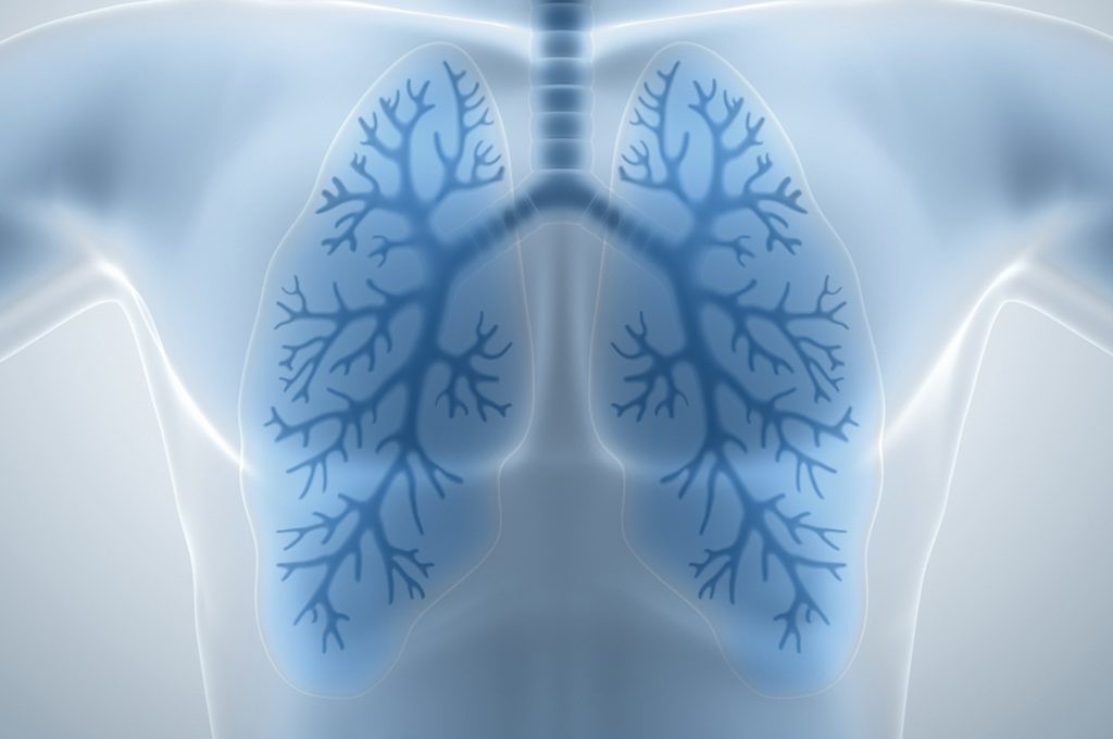
Before gaining a better understanding of what pulmonary hypertension is, it is appropriate to do a little review of the exchanges that the heart and lungs have.
In a normal condition, the blood starts from the right side of the heart and, through the pulmonary arteries, irrigates all the blood vessels of the lungs, until it reaches the capillaries.
It is in these small vessels that the exchange between carbon dioxide and oxygen occurs.
The pulmonary pressure is generally low, so the right side of the heart has less muscularity than the left side (which instead sends blood to all the other parts of the body), which needs more pressure.
Sometimes, however, it happens that due to structural changes in the blood vessels (narrowing, obstruction, parietal thickening) the pressure increases from an average of 14mmHg to 25mmHg.
Under these conditions, the right ventricle is subjected to an excessive load of pressure and volume and could reach contractile failure and thus decompensation.
Or, it could happen that the right ventricle thickens and swells excessively, developing the so-called pulmonary heart, resulting in right heart failure.
If neglected or not properly treated, pulmonary hypertension can even culminate in fatal heart failure.
What are the causes?
To identify the causes of pulmonary hypertension, a distinction must be made in the disease.
It is possible for it to occur in the absence of any particular trigger or previous illness: we speak in this case of primary or idiopathic pulmonary hypertension.
Women – twice as many as men – between 30 and 50 years of age are particularly affected. In this case, the cause is unknown, but as research progresses, some associations with genetic mutations are being identified.
Unfortunately, the mechanism by which these mutations cause pulmonary hypertension is still unknown.
In addition, it has become apparent that the intake of drugs and substances such as fenfluramine (a substance used in weight loss), amphetamines, cocaine and Selective Serotonin Reuptake Inhibitors (SSRIs) can be serious risk factors for developing the disease.
Pulmonary hypertension can also develop in association with other diseases, in this case we speak of acquired or secondary hypertension, which is much more common than the former.
But what are these driving diseases? Emphysema, pulmonary fibrosis, chronic obstructive pulmonary disease and other pulmonary diseases, as well as sleep apnoea, respiratory pathologies linked to sleep disorders.
Still remaining in the lung area, hypertension can be caused by embolisms in the area.
Heart defects or diseases of the left heart can also be a cause, as can autoimmune diseases of the connective tissue, such as scleroderma or lupus erythematosus.
Finally, there are other diseases that can become a trigger for pulmonary hypertension, such as sickle cell anaemia, chronic liver disease and HIV.
Symptoms of pulmonary hypertension
Generally, pulmonary hypertension is manifested by rather abnormal shortness of breath (or dyspnoea), which occurs even during very light physical activity.
Accompanying dyspnoea is an easy loss of energy, chronic fatigue, feeling light-headed, light-headedness under even mild exertion and fainting.
In the more advanced stages of the disease, symptoms worsen: one may have difficulty breathing even at rest, pain very similar to angina pectoris, caused by the suffering of the right heart, and fluid stagnation, resulting in oedema of the lower limbs.
The diagnosis
Obviously, it is not possible to make a self-diagnosis of pulmonary hypertension; if you realise that something is wrong with your health to such an extent that you suspect this pathology, it is always a good idea to consult your general practitioner first, who will direct you to the appropriate specialist.
The case of secondary pulmonary hypertension is different: generally with that type of pathology listed above, one is already being followed by a specialist who will know how to prescribe the right diagnostic tests to best formulate the diagnosis.
Let’s take a step-by-step look at what tests are usually prescribed for a correct diagnosis.
Chest X-ray, which highlights any dilation of the pulmonary arteries.
Transthoracic echocardiography. This provides an accurate view of the heart and any morphological changes in the right atrium and ventricle, which we have seen are a consequence of increased pulmonary pressure. In addition, if an echodoppler is performed, an indirect estimate of the maximum pressure in the pulmonary artery can also be obtained.
Spirometry to detect lung abnormalities. This involves blowing into a tube connected to a device that measures various breathing parameters.
Angio computed tomography of the chest, an X-ray test to observe the pulmonary arteries and detect the presence of occlusions
Pulmonary perfusory scintigraphy, an investigation that allows the blood circulation of the lungs to be photographed in order to observe obstructions or defects in blood supply.
All these tests are non-invasive and are preliminary to the introduction of a catheter into the heart, the only method for a definitive diagnosis.
The catheter will have to start in an arm or leg to reach the right heart and be able to directly measure certain parameters, such as atrium pressure, mean pulmonary pressure and cardiac output.
In addition, only with cardiac catheterisation is it possible to perform the pulmonary vaso-reactivity test: the pulmonary blood vessels are dilated using certain drugs in order to identify any problems in the vessels.
Other tests can also be administered to confirm the diagnosis of pulmonary hypertension, measure its severity and establish its cause:
Blood tests to rule out the presence of autoimmune diseases.
CT angiography to examine for blood clots in the lungs.
EGA, Haemogasanalysis to measure the amount of oxygen and carbon dioxide in the blood through an arterial sampling.
Cardiopulmonary stress test.
Is it possible to prevent pulmonary hypertension?
As far as primary pulmonary hypertension is concerned, it is difficult to think of prevention, other than to advise against taking the substances listed above that could promote the onset of the disease.
Nor is there any real prevention for secondary pulmonary hypertension, other than treating one’s medical condition as best one can to reduce the risk factors that could cause hypertension.
How is pulmonary hypertension treated?
Fortunately, research and medical innovations are progressing from year to year: until recently, the only possible solution to pulmonary hypertension was a lung transplant or, in the case of severe heart impairment, a heart and lung transplant.
Obviously, this was a solution that was only practised in the most severe cases because the risks and contraindications are so many.
Today, however, there are several treatments that do not definitively solve the problem but slow down the progression of the disease and definitely improve the quality of life.
It must be said, however, that in the most extreme cases of patients in whom the progression of hypertension is not stopped, the only solution still remains transplantation.
Obviously, treatment will be easier when there is an exactly identified cause.
Let us now look specifically at what treatments are used in most cases:
- Administration of drugs that manage to vasodilate the pulmonary circulation: calcium antagonists, prostacyclins, anti-endothelin drugs and phosphodiesterase type 5 inhibitors (sildenafil and the like).
- These substances are able to reduce blood pressure in the pulmonary arteries. This can decisively improve the quality of the patient’s everyday life, extend life expectancy and reduce the likelihood of an impending transplant. Generally, vasodilators are tested on the patient during carotid catheterisation, because they may be dangerous in some individuals.
- Administration of oral anticoagulants, which can be combined with diuretics and other heart failure therapies in the event of circulatory decompensation. These drugs may also be prescribed to prevent symptomatic complications. In particular, diuretics are used to ensure that the right ventricle maintains a normal volume and to reduce swelling in the limbs; while anticoagulants, by preventing blood clotting, reduce the risk of pulmonary embolism.
- If reduced blood oxygenation is noted in the patient, oxygen can be administered via nasal cannulae or oxygen masks. The consequence will be to reduce the blood pressure in the pulmonary arteries and relieve shortness of breath.
Obviously, the case of a secondary form of the disease will be different: therapy will mainly be based on treatments to cure the condition.
Read Also
Emergency Live Even More…Live: Download The New Free App Of Your Newspaper For IOS And Android
Management Of The Patient With Acute And Chronic Respiratory Insufficiency: An Overview
Obstructive Sleep Apnoea: What It Is And How To Treat It

Pneumology: Difference Between Type 1 And Type 2 Respiratory Failure
Capnography In Ventilatory Practice: Why Do We Need A Capnograph?
Clinical Review: Acute Respiratory Distress Syndrome
What Is Hypercapnia And How Does It Affect Patient Intervention?
Ventilatory Failure (Hypercapnia): Causes, Symptoms, Diagnosis, Treatment
How To Choose And Use A Pulse Oximeter?
Mild, Severe, Acute Pulmonary Insufficiency: Symptoms And Treatment
Pulmonary Arterial Hypertension: What Is It And Why Is It Important?
Equipment: What Is A Saturation Oximeter (Pulse Oximeter) And What Is It For?
Basic Understanding Of The Pulse Oximeter
Three Everyday Practices To Keep Your Ventilator Patients Safe
Medical Equipment: How To Read A Vital Signs Monitor
Ambulance: What Is An Emergency Aspirator And When Should It Be Used?
Ventilators, All You Need To Know: Difference Between Turbine Based And Compressor Based Ventilators
Life-Saving Techniques And Procedures: PALS VS ACLS, What Are The Significant Differences?
The Purpose Of Suctioning Patients During Sedation
Supplemental Oxygen: Cylinders And Ventilation Supports In The USA
Basic Airway Assessment: An Overview
Ventilator Management: Ventilating The Patient
Emergency Equipment: The Emergency Carry Sheet / VIDEO TUTORIAL
Defibrillator Maintenance: AED And Functional Verification
Respiratory Distress: What Are The Signs Of Respiratory Distress In Newborns?
EDU: Directional Tip Suction Catheter
Suction Unit For Emergency Care, The Solution In A Nutshell: Spencer JET
Airway Management After A Road Accident: An Overview
Tracheal Intubation: When, How And Why To Create An Artificial Airway For The Patient
What Is Transient Tachypnoea Of The Newborn, Or Neonatal Wet Lung Syndrome?
Traumatic Pneumothorax: Symptoms, Diagnosis And Treatment
Diagnosis Of Tension Pneumothorax In The Field: Suction Or Blowing?
Pneumothorax And Pneumomediastinum: Rescuing The Patient With Pulmonary Barotrauma
ABC, ABCD And ABCDE Rule In Emergency Medicine: What The Rescuer Must Do
Multiple Rib Fracture, Flail Chest (Rib Volet) And Pneumothorax: An Overview
Internal Haemorrhage: Definition, Causes, Symptoms, Diagnosis, Severity, Treatment
Assessment Of Ventilation, Respiration, And Oxygenation (Breathing)
Oxygen-Ozone Therapy: For Which Pathologies Is It Indicated?
Difference Between Mechanical Ventilation And Oxygen Therapy
Hyperbaric Oxygen In The Wound Healing Process
Venous Thrombosis: From Symptoms To New Drugs
What Is Intravenous Cannulation (IV)? The 15 Steps Of The Procedure
Nasal Cannula For Oxygen Therapy: What It Is, How It Is Made, When To Use It
Nasal Probe For Oxygen Therapy: What It Is, How It Is Made, When To Use It
Oxygen Reducer: Principle Of Operation, Application
How To Choose Medical Suction Device?
Holter Monitor: How Does It Work And When Is It Needed?
What Is Patient Pressure Management? An Overview
Head Up Tilt Test, How The Test That Investigates The Causes Of Vagal Syncope Works
Cardiac Syncope: What It Is, How It Is Diagnosed And Who It Affects
Cardiac Holter, The Characteristics Of The 24-Hour Electrocardiogram
Stress And Distress During Pregnancy: How To Protect Both Mother And Child
Respiratory Distress: What Are The Signs Of Respiratory Distress In Newborns?
Sepsis: Survey Reveals The Common Killer Most Australians Have Never Heard Of
Sepsis, Why An Infection Is A Danger And A Threat To The Heart
Respiratory Distress Syndrome (ARDS): Therapy, Mechanical Ventilation, Monitoring
Respiratory Assessment In Elderly Patients: Factors To Avoid Respiratory Emergencies