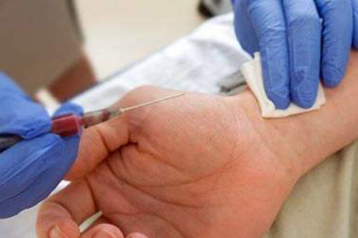
Arterial haemogas analysis: procedure and data interpretation
Haemogasanalysis, often abbreviated to ‘haemogas’, is a diagnostic blood test that consists of measuring the amount of oxygen and carbon dioxide in arterial blood and the pH of the blood
There are two different types of haemogas tests: venous venous and arterial
Arterial haemogas analysis involves taking blood from an artery.
The sampling is more complex for the operator and more uncomfortable for the patient, as the arteries are located deeper so the needle must be inserted deeper to reach them based on knowledge of human anatomy.
Nevertheless, arterial haemogas analysis provides more accurate data on lung function than venous haemogas, as it measures oxygen, carbon dioxide and pH levels, while venous blood analysis is useful for measuring pH in the case of diseases that require metabolic assessments.
Arterial haemogas: what is it?
Systemic arterial haemogas analysis obtains the sample to be analysed via the patient’s radial (wrist) artery or – more rarely – from the brachial (anterior aspect of the elbow) or femoral (groin) artery.
Arterial haemogas is used to measure the amount of oxygen and carbon dioxide in our blood and the pH of the blood.
Arterial blood gas: what is it used for?
Arterial haemogas is useful in all cases where the presence and extent of respiratory insufficiency is to be tested, in combination with other tests.
For the diagnosis of respiratory insufficiency, the doctor also relies on
- Laboratory tests: haemoglobin saturation, haematocrit, urinary output and renal function (azotemia, creatininemia).
- Diagnostic imaging: electrocardiogram, spirometry and other respiratory function tests, echocardiogram, chest X-ray, CT scan, CT angiography, lung scintigraphy.
A blood gas can also be performed to assess the effectiveness of a therapy, in particular the administration of oxygen, or to monitor patients who have been given prolonged anaesthesia during surgery.
Finally, this test is prescribed for patients presenting with sudden respiratory distress (dyspnoea), cyanosis, the onset of abnormal frequent breathing, significant use of accessory respiratory muscles, sudden onset or progression of cardiac arrhythmias, acute hypotension, acute deterioration of neurological function, kidney diseases and metabolic diseases.
Duration of the systemic arterial haemogasanalysis test
Arterial haemogas analysis takes a few minutes, but the patient should not leave the sampling point for at least 10-15 minutes. As a rule, the test result is given at the same sitting as the blood sample (within 30 minutes).
Arterial blood gas preparation rules
To undergo arterial blood gas analysis, fasting is not required, nor is the suspension of any ongoing therapies.
Is arterial blood gas painful?
Needless to lie to you: the test is considered moderately painful.
The patient should report if he or she easily experiences lipotimic episodes (fainting) with blood samples.
The good news is that – if the operator is experienced – it lasts only a few seconds.
In order to alleviate any pain caused by the puncture, an anaesthetic ointment can be applied topically or, alternatively, an infiltration of lidocaine can be performed.
Contraindications of haemogas
Before performing systemic arterial blood gas analysis, the patient must report any medications that interfere with coagulation (TAO).
For patients undergoing oxygen therapy, the therapeutic condition under which the test is to be performed must be indicated: with or without oxygen.
Warnings
Tamponade after blood sampling is much more important than with traditional venous sampling due to the greater pressure of the arteries compared to the veins: after arterial sampling, a tamponade bandage is performed, which should not be removed for more than 1 hour, except in the event of bleeding.
Interpretation of blood gas values
Pa02
PaO2 is the arterial partial pressure of O2 in the blood.
It is expressed in mmHg and the optimal value is between 80 and 100 mmHg.
This value changes with increasing age, so there is a progressive and physiological reduction.
In a young person, Pa02 is normally around 95-100 mmHg in ambient air.
P/F Ratio
The P/F ratio is the ratio of Pa02 to FiO2 and is an indicator of alveolar respiration: P/F = PaO2/Fi02
In a healthy patient, the value is around 450.
A P/F above 350 is considered normal; below 200 is an indication of respiratory failure.
The pH
The pH indicates the acid-base balance. The normal pH value is between 7.35 and 7.45.
If the pH is:
- <7.35, we speak of acidosis
- >7.45 we speak of alkalosis
PaCO2
PaCO2 is the partial pressure of carbon dioxide.
It is measured in mmHg and the optimal value is between 35 and 45 mmHg.
If PaCO2 is:
- <35, we speak of respiratory alkalosis
- >45, we speak of respiratory acidosis
HCO3
HCO3 refers to bicarbonates, the optimal value of which is between 22-26 Mmol/l (millimoles per litre).
If HCO3:
- <22 one speaks of metabolic acidosis
- >26 one speaks of metabolic alkalosis
BE
BE is a parameter assessing base excess.
The reference value is between -2 and +2 mmol/l.
When this value becomes negative, it means that there is a base deficiency and that the patient is in a condition of metabolic acidosis.
This value is used to choose the appropriate treatment for the patient in acidosis.
Electrolytes
The Ega also evaluates electrolytes.
These can also be measured with a normal venous blood sample, but the Ega certainly has the advantage of being more immediate and faster.
In particular, it measures:
- sodium: the optimal value is 135 – 145 mEq/l
- potassium: 3.5 – 5 mEq/l
- Calcium: 8.5 – 10.5 mEq/l
- Chlorine: 95 -105 mEq/l
- Electrolyte control with Ega is particularly important in the dialysed patient.
In fact, dialysis treatment leads to a significant change in blood electrolytes, which is why it is important to carry out checks during treatment to detect abnormalities in good time.
Lactates
Finally, the Ega is able to measure lactates, the normal value of which is < 4 mEq/l.
Lactic acid is produced by cell metabolism; under hypoxic conditions, cells may use less efficient energy production, causing excessive production or poor elimination of lactates.
pH and paCO2 values are closely related.
When tested in combination they provide an indication of the patient’s condition.
Blood gas values and patient condition
Respiratory acidosis (low pH and increased paCO2) is commonly caused by:
- pneumonia;
- COPD;
- depression of respiratory centres secondary to opiate or benzodiazepine intoxication;
- airway obstruction (e.g. PNX).
The patient may present with a low respiratory rate, disoriented or soporific and may complain of headache.
Metabolic acidosis (low pH and low paCO2), on the other hand, is commonly caused by:
- diabetes;
- renal insufficiency;
- alcohol intoxication;
- an abnormal loss of bicarbonate (diarrhoea, vomiting, diabetic ketoacidosis, increased metabolism, prolonged fasting).
The patient is soporific to the point of coma, hyperventilates in order to compensate and may be asthenic.
Respiratory alkalosis (increased pH and decreased paCO2) is caused by:
- severe exercise, hypoxia or anoxia, hyperventilation;
- pain or stress;
- brain trauma;
- damage to the respiratory centre (meningitis, encephalitis);
- fever;
- drug overdose.
The patient is tachypnoic, with altered state of consciousness and may present convulsions.
Metabolic alkalosis (high pH and high paCO2) is caused by:
- protracted vomiting;
- hypokalaemia;
- cirrhosis;
- reabsorption of bicarbonate (use of diuretics, vomiting, sodium retention);
- excessive ingestion of alkali (sodium bicarbonate).
The patient presents bradypnoic and with shallow breathing. He has dizziness, muscle hypertone, is irritable and disoriented.
Arterial haemogas analysis: procedure
- Before carrying out the procedure, the Allen Test should be completed to check the patency of the ulnar artery if the radial artery is chosen as the site for needle insertion.
- The material used can be distinguished as follows
- kit for haemogasanalysis or heparin syringe 10 ml;
- stopper for haemogasanalysis syringe;
- sterile gauze;
- adhesive plaster;
- chlorhexidine disinfection swab;
- biological specimen transport bag;
- appropriate labels for samples;
- container with ice;
- disposable gloves depending on the institution.
At this point we are ready for the procedure:
- Obtain all necessary materials. Check the expiry date of the material. Check the doctor’s order for performing haemogasanalysis. Check the patient’s medical records to make sure that the patient has not been aspirated in the last 15 minutes. If necessary, administer local anaesthetic and wait for it to take effect. Bring the necessary equipment to the patient’s bedside.
- Perform hand hygiene and wear personal protective equipment if indicated. Check the patient’s identification and confirm his or her identity. Tell the patient that it is necessary to draw arterial blood, explaining the procedure. Close the curtains around the bed and close the door to the room if possible. Compare the sample label with the patient.
- Have good lighting. Artificial light is recommended. Place a waste container within reach. If the patient is in bed, he/she should be asked to lie on his/her back with head slightly raised and arms at his/her sides. The outpatient should be asked to sit on a chair and rest his arm on an armrest or table. Place a waterproof towel under the arm and a rolled up towel under the wrist.
- Perform the Allen test before taking a sample from the radial artery. Have the patient close their fist to decrease blood flow to the hand. Using the middle and index finger, press on the radial and ulnar artery. Hold the position for a few seconds.
- Without moving the fingers from the arteries ask the patient to open his fist and hold the hand in a relaxed position. The palm of the patient’s hand should be pale as finger pressure has impeded arterial blood flow.
- Release the pressure on the ulnar artery. If the hand turns pink then blood perfusion fills the vessels and it is safe to perform the radial artery puncture. If, on the other hand, the hand does not turn pink, the Allen test must be performed on the other arm.
- Put on disposable gloves and locate the radial artery, palpating it lightly for a strong pulse. Clean the site with antimicrobial swab. If chlorhexidine is used, perform a forward/backward motion, rubbing for approximately 30 seconds. Allow the skin to dry. After disinfection the site should not be palpated unless sterile gloves are worn.
- Stabilise the hand with the wrist extended over the rolled up towel, palm facing upwards. Palpate the artery over the puncture site with the index and middle finger of the non-dominant hand while holding the syringe with the dominant hand over the puncture site. Do not directly touch the area to be pricked.
- Hold the nozzle of the needle upwards at a 45 degree angle to the radial beat with the syringe parallel to the course of the artery. When puncturing the brachial artery hold the needle at a 60 degree angle.
- Prick the skin and the artery simultaneously. Observe the reflux of blood into the syringe. The pulsating blood will reflux into the syringe. Do not pull the plunger. Fill the syringe to 5 ml.
- After drawing blood, withdraw the syringe while the non-dominant hand begins to compress the arterial puncture site with the 5×5 gauze. Compress strongly until the blood flow stops or for at least 5 minutes. If the patient is on anticoagulant therapy or has a blood dyscrasia, apply pressure for 10-15 minutes. If necessary, ask a support assistant to hold the gauze in place while you prepare the sample for transport to the lab but never ask the patient to hold the gauze.
- When bleeding stops and a reasonable amount of time has passed, apply an adhesive bandage or small compression dressing. Once the sample has been obtained, check for any air bubbles. If any are present, remove them by holding the syringe in an upright position and slowly expelling some blood onto gauze.
- Insert the needle guard. Place the airtight cap on the tip of the syringe. Gently rotate the syringe to ensure good distribution of the heparin. Do not shake. Place the syringe in a cup or bag filled with ice.
- Place the label on the syringe according to the institution’s instructions. Place the syringe immersed in ice in a biohazard bag. Discard the needle in the sharps container and perform hand hygiene.
- Take the sample immediately to the laboratory.
Read Also
Emergency Live Even More…Live: Download The New Free App Of Your Newspaper For IOS And Android
Pulse Oximeter Or Saturimeter: Some Information For The Citizen
Oxygen Saturation: Normal And Pathological Values In The Elderly And Children
Equipment: What Is A Saturation Oximeter (Pulse Oximeter) And What Is It For?
How To Choose And Use A Pulse Oximeter?
Basic Understanding Of The Pulse Oximeter
Capnography In Ventilatory Practice: Why Do We Need A Capnograph?
Clinical Review: Acute Respiratory Distress Syndrome
What Is Hypercapnia And How Does It Affect Patient Intervention?
Ventilatory Failure (Hypercapnia): Causes, Symptoms, Diagnosis, Treatment
How To Choose And Use A Pulse Oximeter?
Equipment: What Is A Saturation Oximeter (Pulse Oximeter) And What Is It For?
Basic Understanding Of The Pulse Oximeter
Three Everyday Practices To Keep Your Ventilator Patients Safe
Medical Equipment: How To Read A Vital Signs Monitor
Ambulance: What Is An Emergency Aspirator And When Should It Be Used?
Ventilators, All You Need To Know: Difference Between Turbine Based And Compressor Based Ventilators
Life-Saving Techniques And Procedures: PALS VS ACLS, What Are The Significant Differences?
The Purpose Of Suctioning Patients During Sedation
Supplemental Oxygen: Cylinders And Ventilation Supports In The USA
Basic Airway Assessment: An Overview
Ventilator Management: Ventilating The Patient
Emergency Equipment: The Emergency Carry Sheet / VIDEO TUTORIAL
Defibrillator Maintenance: AED And Functional Verification
Respiratory Distress: What Are The Signs Of Respiratory Distress In Newborns?
EDU: Directional Tip Suction Catheter
Suction Unit For Emergency Care, The Solution In A Nutshell: Spencer JET
Airway Management After A Road Accident: An Overview
Tracheal Intubation: When, How And Why To Create An Artificial Airway For The Patient
What Is Transient Tachypnoea Of The Newborn, Or Neonatal Wet Lung Syndrome?
Traumatic Pneumothorax: Symptoms, Diagnosis And Treatment
Diagnosis Of Tension Pneumothorax In The Field: Suction Or Blowing?
Pneumothorax And Pneumomediastinum: Rescuing The Patient With Pulmonary Barotrauma
ABC, ABCD And ABCDE Rule In Emergency Medicine: What The Rescuer Must Do
Multiple Rib Fracture, Flail Chest (Rib Volet) And Pneumothorax: An Overview
Internal Haemorrhage: Definition, Causes, Symptoms, Diagnosis, Severity, Treatment
Assessment Of Ventilation, Respiration, And Oxygenation (Breathing)
Oxygen-Ozone Therapy: For Which Pathologies Is It Indicated?
Difference Between Mechanical Ventilation And Oxygen Therapy
Hyperbaric Oxygen In The Wound Healing Process
Venous Thrombosis: From Symptoms To New Drugs
What Is Intravenous Cannulation (IV)? The 15 Steps Of The Procedure
Nasal Cannula For Oxygen Therapy: What It Is, How It Is Made, When To Use It
Nasal Probe For Oxygen Therapy: What It Is, How It Is Made, When To Use It
Oxygen Reducer: Principle Of Operation, Application
How To Choose Medical Suction Device?
Holter Monitor: How Does It Work And When Is It Needed?
What Is Patient Pressure Management? An Overview
Head Up Tilt Test, How The Test That Investigates The Causes Of Vagal Syncope Works
Cardiac Syncope: What It Is, How It Is Diagnosed And Who It Affects
Cardiac Holter, The Characteristics Of The 24-Hour Electrocardiogram
Stress And Distress During Pregnancy: How To Protect Both Mother And Child
Respiratory Distress: What Are The Signs Of Respiratory Distress In Newborns?
Sepsis: Survey Reveals The Common Killer Most Australians Have Never Heard Of
Sepsis, Why An Infection Is A Danger And A Threat To The Heart
Respiratory Distress Syndrome (ARDS): Therapy, Mechanical Ventilation, Monitoring
Respiratory Assessment In Elderly Patients: Factors To Avoid Respiratory Emergencies
Alterations In Acid-Base Balance: Respiratory And Metabolic Acidosis And Alkalosis
Management Of The Patient With Acute And Chronic Respiratory Insufficiency: An Overview
Obstructive Sleep Apnoea: What It Is And How To Treat It
Pneumology: Difference Between Type 1 And Type 2 Respiratory Failure
Ventilatory Management Of The Patient: Difference Between Type 1 And Type 2 Respiratory Failure


