
Electrocardiogram, an overview
The electrocardiogram, or ECG, is an instrumental diagnostic test that uses an electrocardiograph to record and graphically reproduce the heart’s electrical activity through a series of electrodes
By monitoring the pumping activity of the heart, i.e. contractions and relaxations, it is possible to detect probable heart disease, arrhythmias, myocardial infarction, abnormality of the atrium or ventricle of the heart, coronary artery disease, etc.
The electrocardiogram can be used to assess the proper functioning of implantable pacemakers or defibrillators in those who need them to normalise cardiac rhythm.
There are three types of electrocardiogram: resting ECG, dynamic Holter ECG and exercise ECG
Through the electrocardiographic tracing, the cardiologist is able to understand the health status and functioning of the heart.
If you are on medication or have a pacemaker and the like, you should mention this to your cardiologist.
Generally, in a tracing lines describe rhythm and activity of the heart, in medical terms they are called waves; the distance between the waves and their appearance allow the cardiologist to read them and consequently understand the state of health of the heart.
Resting Electrocardiogram (Resting ECG)
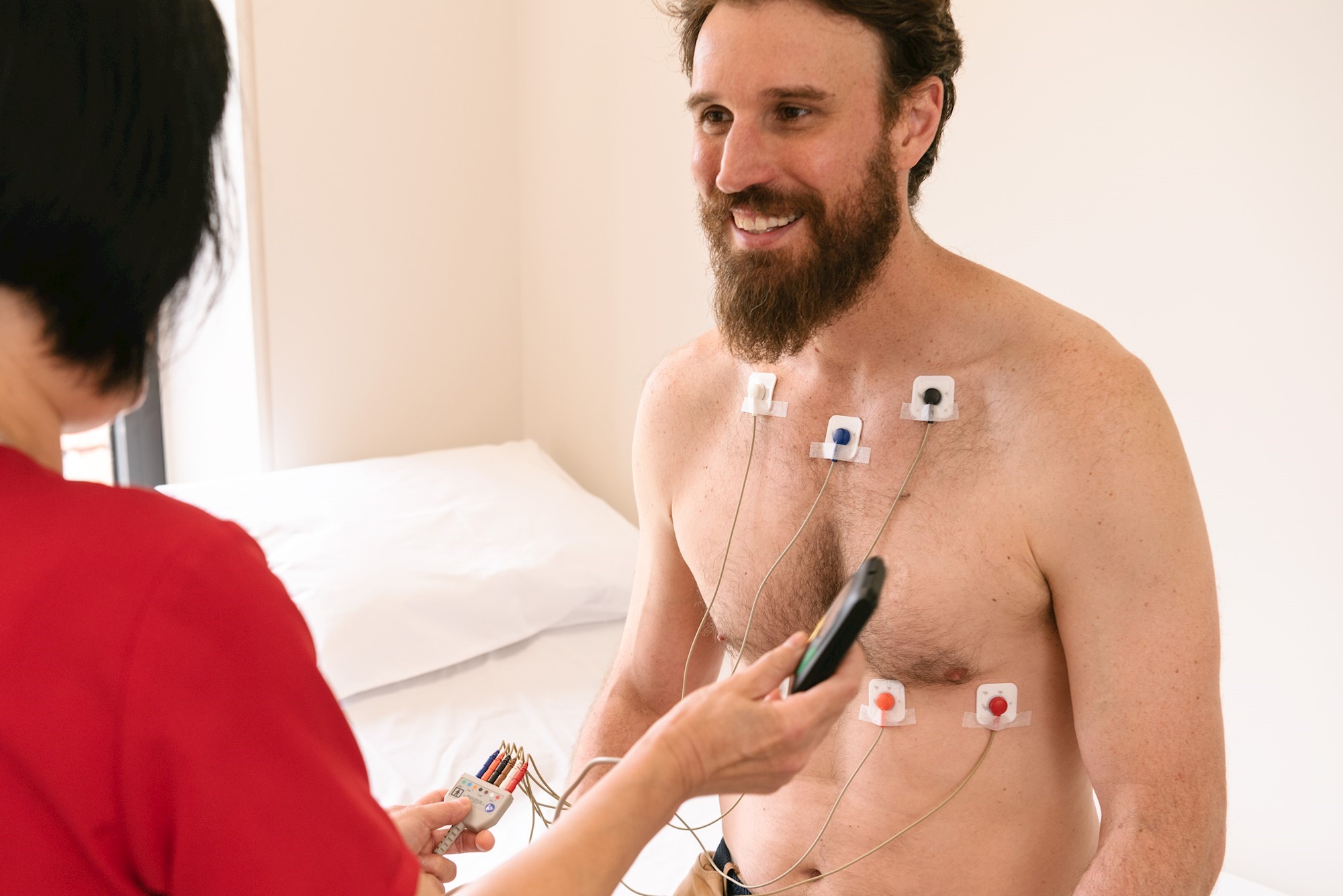
After we sit on the couch, the electrodes of the electrocardiograph are applied to our chest, arms and legs.
The electrodes are metal plates that can be applied by means of an adhesive part, suction cups or adhesive gel.
Once the electrodes have been applied, the electrocardiograph will be started and recording will begin. The recording lasts a few seconds, just long enough to obtain a trace to assess heart function.
During the procedure, the patient should breathe normally but should avoid moving in order not to distort the test results.
The duration of the resting ECG is a few minutes.
Dynamic Holter electrocardiogram
The Holter electrocardiogram uses a portable electrocardiograph to monitor cardiac activity over a period of 24 to 48 hours.
The creation of this portable electrocardiograph stems from the need to capture discontinuous and sporadic cardiac arrhythmias that cannot be detected in the resting ECG.
The electrodes, in this case, are applied only on the chest and are metal plates with an adhesive portion.
The Holter electrocardiogram can be divided into two phases:
- The phase of recording the rhythm and electrical activity of the heart; it is the first phase, running from the installation of the portable electrocardiograph until its removal. The device records and saves the patient’s heart function in an internal memory.
- The second and last phase concerns the graphic translation of what was recorded in the first phase, the trace is created.
A nurse will extrapolate the data recorded by the electrocardiograph using a specific computerised device, while the cardiologist will interpret the trace.
During the recording phase, the patient can perform the usual daily actions, taking care, however, not to detach the electrodes and not to bump the device.
Electrocardiogram Under Stress
The stress electrocardiogram records the cardiac activity of an individual while performing exercise at a certain intensity or, in rarer cases, after taking medication that has the same effects on the heart as exercise.
The purpose of this type of electrocardiogram is to see how the heart behaves when subjected to physical exertion: how the heart rhythm varies, what cardiac problems the body’s demand for more blood can cause.
The application area of the electrodes is only on the thoracic area since placing them in other areas of the body would prevent uninhibited movement during exercise.
The latter mainly consists of pedalling on an exercise bike or walking/running on a treadmill.
The electrocardiogram is a non-invasive and safe procedure, the only drawback being a slight reddening or swelling of the skin in the area where the electrodes were applied.
Should there be any cardiac complications during this type of electrocardiogram, the cause is being under stress and not the electrocardiogram.
Thanks to the electrocardiogram, it is possible to accurately detect alterations in the heart rhythm that may arise due to altered conduction of the nerve impulse through the myocardium or as a result of heart disease, myocardial infarction or cardiomyopathy.
The electrocardiographic tracing of a healthy person consists of five waves, which are denoted by the letters P, Q, R, S and T.
The P wave indicates cardiac atrial contraction; it lasts approximately 0.08 seconds, with a tolerance ranging from 0.05 to 0.12.
After the P wave, there is a straight line that ends at the Q, R, and S waves and is called the PR interval, which lasts from 0.16 to 0.2 seconds.
The Q, R, and S waves form the QRS complex, which represents the contraction of the ventricles and lasts approximately 0.12 seconds. With the contraction of the ventricles we have atrial relaxation.
The T wave: expresses the relaxation of the ventricles.
After the T wave there is again a horizontal stretch ending with a P wave, which represents a new phase of depolarisation and repolarisation of the atria and ventricles, i.e. when the ventricles have to undergo an electrical change to prepare for the next heartbeat.
The P, Q, R, S and T waves together form the PQRST complex. The interval between two PQRST complexes is called the R-R interval, an interval that corresponds to one cardiac cycle.
Read Also
Emergency Live Even More…Live: Download The New Free App Of Your Newspaper For IOS And Android
Heart Pacemaker: How Does It Work?
Cardiac Electrostimulation: The Leadless Pacemaker
Diagnosis Of Mitral Stenosis? Here’s What’s Happening
Inflammations Of The Heart: Myocarditis

Paediatric Pacemaker: Functions And Peculiarities
What Is The Difference Between Pacemaker And Subcutaneous Defibrillator?
Heart: What Is Brugada Syndrome And What Are The Symptoms
Genetic Heart Disease: Brugada Syndrome
Cardiac Arrest Defeated By A Software? Brugada Syndrome Is Near To An End
Heart: Brugada Syndrome And The Risk Of Arrhythmia
Heart Disease: First Study On Brugada Syndrome In Children Under 12 From Italy
Mitral Insufficiency: What It Is And How To Treat It
Semeiotics Of The Heart: History In The Complete Cardiac Physical Examination
Electrical Cardioversion: What It Is, When It Saves A Life
Heart Murmur: What Is It And What Are The Symptoms?
Performing The Cardiovascular Objective Examination: The Guide
Branch Block: The Causes And Consequences To Take Into Account
Cardiopulmonary Resuscitation Manoeuvres: Management Of The LUCAS Chest Compressor
Supraventricular Tachycardia: Definition, Diagnosis, Treatment, And Prognosis
Identifying Tachycardias: What It Is, What It Causes And How To Intervene On A Tachycardia
Myocardial Infarction: Causes, Symptoms, Diagnosis And Treatment
Aortic Insufficiency: Causes, Symptoms, Diagnosis And Treatment Of Aortic Regurgitation
Congenital Heart Disease: What Is Aortic Bicuspidia?
Atrial Fibrillation: Definition, Causes, Symptoms, Diagnosis And Treatment
Ventricular Fibrillation Is One Of The Most Serious Cardiac Arrhythmias: Let’s Find Out About It
Atrial Flutter: Definition, Causes, Symptoms, Diagnosis And Treatment
What Is Echocolordoppler Of The Supra-Aortic Trunks (Carotids)?
What Is The Loop Recorder? Discovering Home Telemetry
Cardiac Holter, The Characteristics Of The 24-Hour Electrocardiogram
Peripheral Arteriopathy: Symptoms And Diagnosis
Endocavitary Electrophysiological Study: What Does This Examination Consist Of?
Cardiac Catheterisation, What Is This Examination?
Echo Doppler: What It Is And What It Is For
Transesophageal Echocardiogram: What Does It Consist Of?
Paediatric Echocardiogram: Definition And Use
Heart Diseases And Alarm Bells: Angina Pectoris
Fakes That Are Close To Our Hearts: Heart Disease And False Myths
Sleep Apnoea And Cardiovascular Disease: Correlation Between Sleep And Heart
Myocardiopathy: What Is It And How To Treat It?
Venous Thrombosis: From Symptoms To New Drugs
Cyanogenic Congenital Heart Disease: Transposition Of The Great Arteries
Heart Rate: What Is Bradycardia?
Consequences Of Chest Trauma: Focus On Cardiac Contusion