
High cervical spine traumas in children: what they are, how to intervene
High cervical spine traumas (C0-C1-C2) are caused by falls or road accidents, especially in children under 8 years of age. They may require immobilisation or surgery, even complex surgery
Any traumatic event (accidental fall, collision, sports accident) that involves the bony structures, ligaments, blood vessels or nerve structures of the neck, and alters their normal function, more or less severely, is defined as cervical trauma.
WOULD YOU LIKE TO GET TO KNOW RADIOEMS? VISIT THE RADIOEMS RESCUE BOOTH AT EMERGENCY EXPO
Cervical spine trauma in paediatric age can be divided into 2 main groups:
- Traumas involving the high or axial cervical spine (C0-C1-C2 vertebrae);
- Traumas involving the low or subaxial cervical spine (C3 to C7 vertebrae).
Traumas of the high cervical spine are more frequent in younger children, usually under 8 years of age.
They are more susceptible for several reasons:
- Lower holding force (laxity) of the ligaments that stabilise the cervical vertebrae between them;
- Predominantly cartilaginous (therefore less solid) structure of the joints and a shape of the vertebrae that ‘predisposes’ them to greater instability than those of adults;
- Larger head size compared to the adult body;
- High cervical fractures account for less than 1% of all paediatric fractures. They are mainly caused by sports injuries in younger children and by bicycle or motorbike accidents in adolescents.
The main and most frequent consequences of high cervical spine trauma can be distinguished into:
- Bone fractures (C0-C1-C2);
- Fractures of the tooth of the C2 vertebra also known as the epistrophe (fractures of the epistrophe tooth);
- C1-C2 dislocation/subluxation.
Fractures can involve different parts of the vertebrae:
- The most frequent are fractures of the epistropheus tooth (C2), which are frequent in children under 7 years of age and are often caused by falls from heights or road accidents involving children secured in a car seat with the front end facing in the direction of travel (trauma from abrupt deceleration);
- fractures of the arch of the atlas (C1);
- Injuries also involving the occipital bone are rarer (C0).
C1-C2 subluxation is an alteration of the normal rotational relationships between the atlas (C1) and the epistrophe (C2).
Subluxation can have different levels of severity (1 to 4), levels that require various types of treatment, either conservative or surgical.
Dislocation, on the other hand, is the loss of the normal anatomical relationship between C1 and C2 with potentially very serious neurological consequences if not acted upon promptly.
Dislocation most frequently benefits from prolonged surgical/conservative treatment.
With regard to clinical manifestations, typical is an intense and painful contracture of the neck muscles causing a ‘torticollis’, or an attitude of the child with the head flexed forward and a tendency, in less severe cases, to protect himself with his hands in lateral head movements.
Neurological alterations with partial impairment of arm and leg movement or symptoms such as loss of consciousness are more serious indicators.
High cervical spine trauma, from diagnosis in the emergency room to damage assessment
Diagnosis is based on an examination of the patient generally carried out in the emergency room, with assessment of any vascular or neurological damage.
Fundamental to the diagnosis is the acquisition of X-ray images taken in precise projections according to the clinical suspicion.
Computed tomography and magnetic resonance imaging complete the picture and make it possible, through precise computer measurements, to make a diagnosis of instability with subluxation or dislocation, or fracture.
The clinical assessment is important, as the possibility of a false positive (i.e. apparent subluxation, but in reality it is lateral positioning of the head at the time of acquisition of the above-mentioned X-ray image) is frequent.
Treatment can be conservative or surgical. In both cases, reduction manoeuvres, with or without anaesthesia, may be necessary to allow the correct repositioning of C1 and C2 in order to restore the normal relationship of the joint between these two vertebrae.
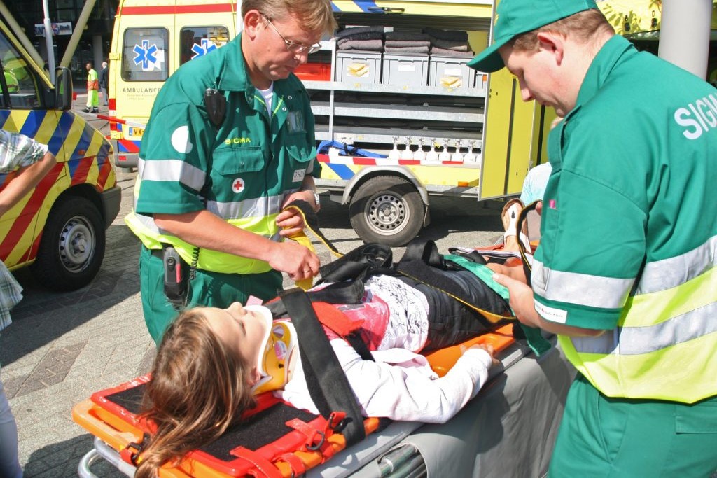
Conservative treatment involves the use of immobilisation collars for various periods of time, depending on the extent of the injury, together with the use of anti-inflammatory, pain-relieving and muscle-relaxing drugs.
Collars can be soft (e.g. Schanz collar) or rigid (e.g. Philadelphia collar) and must be worn continuously for different lengths of time depending on the fracture breakdown or the reducibility and maintenance in correction of the dislocation/subluxation.
Collars allow the healing of injured structures by immobilising the movements in which they are involved.

Surgical treatments for high cervical spine trauma may mainly include:
- Halo transcranial traction placement: a metal ring (halo) is applied around the head under general anaesthesia, attaching the ring to the child’s skull with multiple nails. The halo is not painful and is generally well tolerated. Weights are attached to the ring, which keep the unstable vertebrae spaced out and locked (in reduction) by pulling the head in relation to the spine. It may take several weeks before this device is safely removed;
- Placement of screws (or hooks) and bars to fix unstable or fractured vertebrae to adjacent ‘healthy’ vertebrae in the correct position. This intervention is called ‘stabilisation’ which can be temporary or definitive. If definitive, it is more properly called ‘arthrodesis’. This intervention is reserved for cases of severe instability, with or without decomposed fractures, dislocation that persists despite repeated attempts with the other treatments described, especially when there are serious neurological risks for young patients.
These treatments require frequent clinical and radiographic follow-up to assess the progress of the instability and the healing of the fracture.
Removal of the collars or halo traction usually occurs no sooner than 4-6 weeks, but may occur at 3 months in severe cases.
Read Also
Emergency Live Even More…Live: Download The New Free App Of Your Newspaper For IOS And Android
KED Extrication Device For Trauma Extraction: What It Is And How To Use It
What Should Be In A Paediatric First Aid Kit
Does The Recovery Position In First Aid Actually Work?
Is Applying Or Removing A Cervical Collar Dangerous?
Cervical Collars : 1-Piece Or 2-Piece Device?
Cervical Collar In Trauma Patients In Emergency Medicine: When To Use It, Why It Is Important
What Is Traumatic Brain Injury (TBI)?
Pathophysiology Of Thoracic Trauma: Injuries To The Heart, Great Vessels And Diaphragm
Cardiopulmonary Resuscitation Manoeuvres: Management Of The LUCAS Chest Compressor
Chest Trauma: Clinical Aspects, Therapy, Airway And Ventilatory Assistance
Precordial Chest Punch: Meaning, When To Do It, Guidelines
Ambu Bag, Salvation For Patients With Lack Of Breathing
Blind Insertion Airway Devices (BIAD’s)
UK / Emergency Room, Paediatric Intubation: The Procedure With A Child In Serious Condition
How Long Does Brain Activity Last After Cardiac Arrest?
Quick And Dirty Guide To Chest Trauma
Cardiac Arrest: Why Is Airway Management Important During CPR?