
Cutaneous manifestations of bacterial endocarditis: Osler nodes and Janeway's lesions
Osler nodes and Janeway lesions are two rare but well-known skin manifestations of bacterial endocarditis. They have also rarely been described in systemic lupus erythematosus (SLE), gonococcaemia (gonorrhoea), haemolytic anaemia and typhoid fever
They are important as they may help in the earlier diagnosis of a serious medical disorder.
What is bacterial endocarditis?
Bacterial endocarditis is an infection of the lining of the heart caused by various bacteria.
It most often affects the heart valves.
The bacteria gain access to the heart via the bloodstream; an infection elsewhere in the body may or may not be apparent.
While some bacteria can cause infection in normal heart valves, bacterial endocarditis more commonly affects patients with abnormal valves as a result of previous rheumatic fever, valve surgery/replacement, or congenital abnormalities.
The responsible bacteria include various species of staphylococcus, streptococcus, pseudomonas, bartonella and several other organisms.
Bacterial endocarditis is often divided into ‘acute’ and ‘subacute’ varieties depending on the speed that it progresses.
The symptoms may include fever, lethargy, shortness of breath, chest pain or palpitations.
These symptoms require prompt assessment and investigation by a physician.
Splinter haemorrhage in the proximal nail plate is also a sign of bacterial endocarditis.
FIRST AID: VISIT THE DMC DINAS MEDICAL CONSULTANTS BOOTH AT EMERGENCY EXPO
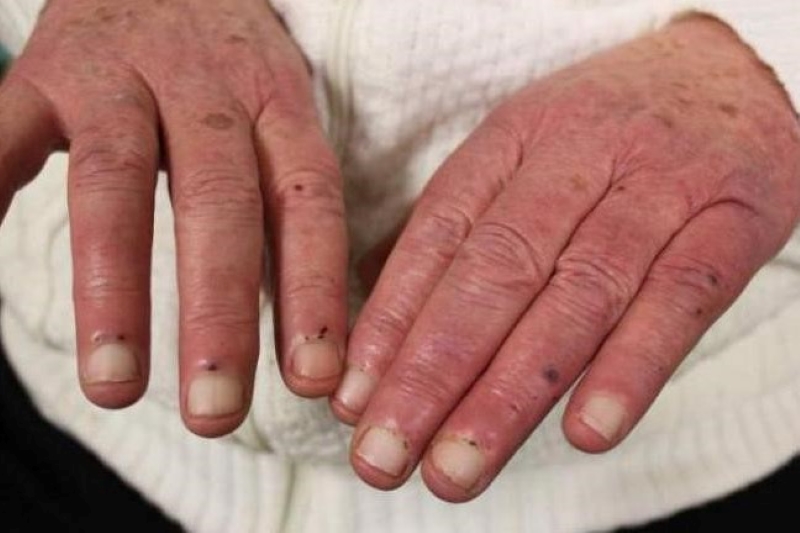
Osler nodes
Osler nodes are red-purple, slightly raised, tender lumps, often with a pale centre.
Pain often precedes the development of the visible lesion by up to 24 hours.
They are typically found on the fingers and/or toes.
They can occur at any time during the course of endocarditis (usually subacute) and last from hours to several days.
How did Osler nodes get their name?
The lesions were first described by French Physicians as ‘Nodosites Cutanees Ephemeres’ meaning ‘Cutaneous nodules of short duration’ and by Dr Mullen of Hamilton.
Parkes Weber later suggested that they are known as Osler nodes in recognition of the fact that Sir William Osler (1849-1919) had “first called attention to their full diagnostic importance”.
His first description of these lesions was in 1893.
Sir William Osler, a Canadian-born Physician, wrote 1344 publications on a wide range of medical topics.
DEFIBRILLATORS: VISIT THE PROGETTI MEDICAL EQUIPMENT SOLUTIONS BOOTH AT EMERGENCY EXPO
What is the cause of Osler nodes?
The underlying cause of the nodes has been debated since Osler first proposed micro-embolisation as a cause (this is the scattering of tiny particles around the bloodstream).
Early reports favoured an allergic or immunological cause, but more recent reports have isolated bacteria from within the nodules.
A skin biopsy (histology) may reveal a neutrophilic vasculitis (inflammation of blood vessels) affecting the glomus apparatus of the ends of the fingers, or microabscess formation without evidence of vasculitis.
It has been postulated that early biopsies show bacteria within the microabscesses and as time progresses, the nodes become sterile and hypersensitivity vasculitis or small vessel vasculitis develops, mediated by the immune system.
What tests should be done?
A careful search for endocarditis is made.
This includes multiple blood cultures, other blood tests, urine tests, ECG, chest X-ray, and an echocardiogram (heart ultrasound scan).
The diagnosis may be elusive.
A skin biopsy may be helpful to confirm the diagnosis of the Osler nodes.
What is the treatment of Osler nodes?
Treatment of Osler nodes is aimed at the bacterial endocarditis and involves intravenous antibiotics and sometimes valve surgery.
The skin lesions tend to heal spontaneously without scarring.
Janeway lesions
In contrast to Osler nodes, Janeway lesions are non-tender, often haemorrhagic (bleeding into the skin), and occur mostly on the palms and soles on the thenar and hypothenar eminences (at the base of the thumb and little finger respectively).
They tend to last days to weeks before healing totally. Janeway lesions are more commonly seen in acute endocarditis, when bacteria such as Staphylococcus aureus may be cultured from them.
The histology is usually consistent with septic micro-embolism (i.e. bacteria may be found within the blood vessels).
References
- Alpert JS, et al. Pathogenesis of Osler nodes. Annals of Internal Medicine 1976;85471–3. PubMed
- Botella R, et al. Janeway lesions differential diagnosis with Osler nodes. Int J Derm 1993;32(9)673–4. PubMed
- Cardullo AC, et al. Janeway lesions and Osler nodes a review of histopathologic findings. J Am Acad Derm 1990;22:1088–90. PubMed
- Freedberg IM, et al. Fitzpatrick’s Dermatology in general medicine. 6th ed. New York, McGraw Hill, 2003.
- Infective Endocarditis – Medscape Reference
- Heart conditions – endocarditis Better Health Channel, Victorian Government (Australia)
Read Also
Emergency Live Even More…Live: Download The New Free App Of Your Newspaper For IOS And Android
Bacterial Endocarditis: Prophylaxis In Children And Adults
Sinus Tachycardia: What It Is And How To Treat It
Defibrillator: What It Is, How It Works, Price, Voltage, Manual And External
The Patient’s ECG: How To Read An Electrocardiogram In A Simple Way

Signs And Symptoms Of Sudden Cardiac Arrest: How To Tell If Someone Needs CPR
Inflammations Of The Heart: Myocarditis, Infective Endocarditis And Pericarditis
Quickly Finding – And Treating – The Cause Of A Stroke May Prevent More: New Guidelines
Atrial Fibrillation: Symptoms To Watch Out For
Wolff-Parkinson-White Syndrome: What It Is And How To Treat It
Do You Have Episodes Of Sudden Tachycardia? You May Suffer From Wolff-Parkinson-White Syndrome (WPW)
Transient Tachypnoea Of The Newborn: Overview Of Neonatal Wet Lung Syndrome
Tachycardia: Is There A Risk Of Arrhythmia? What Differences Exist Between The Two?
Diseases Of The Heart: Postural Orthostatic Tachycardia (POTS)
Supraventricular Tachycardia: Definition, Diagnosis, Treatment, And Prognosis
Identifying Tachycardias: What It Is, What It Causes And How To Intervene On A Tachycardia
Who Can Use The Defibrillator? Some Information For Citizens
Defibrillator Maintenance: What To Do To Comply
Defibrillators: What Is The Right Position For AED Pads?
When To Use The Defibrillator? Let’s Discover The Shockable Rhythms
What Is The Difference Between Pacemaker And Subcutaneous Defibrillator?
What Is An Implantable Defibrillator (ICD)?
What Is A Cardioverter? Implantable Defibrillator Overview
Paediatric Pacemaker: Functions And Peculiarities
Cardiac Arrest: Why Is Airway Management Important During CPR?
Tachycardia: Is There A Risk Of Arrhythmia? What Differences Exist Between The Two?
Do You Have Episodes Of Sudden Tachycardia? You May Suffer From Wolff-Parkinson-White Syndrome (WPW)
Transient Tachypnoea Of The Newborn: Overview Of Neonatal Wet Lung Syndrome
Paediatric Toxicological Emergencies: Medical Intervention In Cases Of Paediatric Poisoning
Valvulopathies: Examining Heart Valve Problems
What Is The Difference Between Pacemaker And Subcutaneous Defibrillator?
Heart Disease: What Is Cardiomyopathy?
Inflammations Of The Heart: Myocarditis, Infective Endocarditis And Pericarditis
Heart Murmurs: What It Is And When To Be Concerned
Broken Heart Syndrome Is On The Rise: We Know Takotsubo Cardiomyopathy
Cardiomyopathies: What They Are And What Are The Treatments
Alcoholic And Arrhythmogenic Right Ventricular Cardiomyopathy
Difference Between Spontaneous, Electrical And Pharmacological Cardioversion
What Is Takotsubo Cardiomyopathy (Broken Heart Syndrome)?
Dilated Cardiomyopathy: What It Is, What Causes It And How It Is Treated
Heart Pacemaker: How Does It Work?
Basic Airway Assessment: An Overview
Assessment Of Abdominal Trauma: Inspection, Auscultation And Palpation Of The Patient
Pain Assessment: Which Parameters And Scales To Use When Rescuing And Treating A Patient
Airway Management After A Road Accident: An Overview
Tracheal Intubation: When, How And Why To Create An Artificial Airway For The Patient
What Is Traumatic Brain Injury (TBI)?
Acute Abdomen: Meaning, History, Diagnosis And Treatment
Poison Mushroom Poisoning: What To Do? How Does Poisoning Manifest Itself?
Chest Trauma: Clinical Aspects, Therapy, Airway And Ventilatory Assistance
The Quick And Dirty Guide To Pediatric Assessment
EMS: Pediatric SVT (Supraventricular Tachycardia) Vs Sinus Tachycardia